| Journal of Clinical Question. 2025; 2(4): e83 https://doi.org/10.69854/jcq.2025.0026 Advance access publication date 22 August 2025 |
 |
Case Report
Takayasu Arteritis with Multivessel Occlusion: Staged Revascularization and Cerebral Perfusion Management: A Case Report
Zaiqiang Yu , Kenyou Murata, Yoshiaki Saito, Tomonori Kawamura, Masahito Minakawa*
Department of Thoracic and Cardiovascular Surgery, Hirosaki University Graduate School of Medicine.
*Corresponding Author: e-mail: masahito@hirosaki-u.ac.jp
Submitted: July 28, 2025 Accepted: August 21, 2025
Clinical Question Box
How can perioperative management reduce the risk of cerebral hyperperfusion syndrome in Takayasu arteritis surgery involving supraaortic trunk and peripheral vessels?
A staged revascularization strategy, combined with cerebral perfusion assessment and meticulous perioperative blood pressure control, is crucial for reducing the risk of hyperperfusion in Takayasu arteritis surgery. Preoperative perfusion imaging of supraaortic trunks and cerebral vessels aids in surgical planning, while careful hemodynamic monitoring helps prevent neurological complications.
Abstract
Background: Takayasu arteritis is a rare large-vessel vasculitis that typically affects young women, but it is also diagnosed in older patients with severe vascular occlusions. Case Presentation: We report a case of a 67-year-old female with Takayasu arteritis, presenting with extensive multivessel occlusions involving the brachiocephalic, carotid, subclavian, mesenteric, and iliac arteries, resulting in severe bilateral limb ischemia. Due to persistent rest pain in both limbs, surgical management involved multibranch revascularization using a prosthetic graft from the brachiocephalic artery to both the axillary and femoral arteries, along with a bypass to the right common carotid artery. Postoperatively, the patient developed cerebral hyperperfusion syndrome complicated by intracerebral hemorrhage, necessitating surgical flow reduction of the carotid bypass. The patient recovered with resolution of limb ischemia and no persistent neurological deficits. Conclusion: This case underscores the complexities of surgical management in Takayasu arteritis, highlighting the critical role of staged revascularization, thorough cerebral perfusion evaluation, and careful perioperative blood pressure control in minimizing hyperperfusion-related risks.
Keywords: Takayasu arteritis, aortitis syndrome, revascularization, cerebral hemorrhage, cerebral hyperperfusion syndrome, case report.
Introduction
Aortitis is a histopathologic diagnosis characterized by inflammation of the aorta. It represents a group of large-vessel diseases with diverse or unknown causes.1 Although rare, aortitis is diagnosed in approximately 1–3 cases per million people annually.2 Immune-mediated conditions that can affect the aorta include Takayasu arteritis, giant cell arteritis, polyarteritis nodosa, Behçet disease, Cogan syndrome, sarcoidosis, spondyloarthropathy, serum sickness, cryoglobulinemia, systemic lupus erythematosus, rheumatoid arthritis, Henoch–Schönlein purpura, and immune complex diseases related to infections or drugs.3
Takayasu arteritis is a particularly rare form of large-vessel vasculitis that predominantly affects young women. It typically presents with symptoms of arterial occlusion affecting the aorta, aortic arch, and major branches, with imaging being essential for evaluation given the complexity of the vascular territories involved.4,5 The condition is also known as pulseless disease or aortic arch syndrome, reflecting its clinical manifestations.6 Although first described by Japanese ophthalmologist Mikoto Takayasu, epidemiological data do not indicate a higher incidence of the disease in Japan.7 The reported incidence ranges from 0.3 to 3.4 cases per million people annually across different countries.8–10 Patients often present with severe limb ischemia and carotid artery occlusion. Management often requires surgical intervention, such as vascular reconstruction with graft replacement or bypass procedures. Endovascular therapy is also utilized in select cases. We present a case of aortitis syndrome treated with multibranch revascularization using a graft extending from the brachiocephalic artery to the limbs and the right carotid artery.
Case Presentation
A 67-year-old female with a history of hypertension was referred to our hospital for evaluation of severe limb ischemia. She had no other significant past medical history. On examination, peripheral pulses were absent in all limbs except for the right femoral artery. The ankle–brachial index (ABI) was 0.66 on the right and 0.56 on the left, findings consistent with severe bilateral limb ischemia accompanied by rest pain. Transthoracic echocardiography revealed good left ventricular function with mild aortic valve regurgitation. Laboratory tests showed a normal white blood cell count (10,600/μL), normal C-reactive protein levels (<0.02 mg/dL), and normal renal function (creatinine 0.49 mg/dL).
Computed tomography angiography (CTA) revealed occlusion of the brachiocephalic and bilateral subclavian arteries, with collateral vessels supplying both the axillary and subclavian arteries. Occlusion of the celiac artery ostium and stenosis of the superior mesenteric and left renal artery ostia were also noted, secondary to renal artery stenosis. The likely mechanism of hypertension is primary hypertension, not related to secondary causes such as renal artery stenosis. A heavily calcified lesion extending from the terminal abdominal aorta to both the common iliac arteries was responsible for the severe limb ischemia (Figs. 1A, 1B). Magnetic resonance demonstrated occlusion of the right common carotid artery and stenosis of the left internal carotid artery (Figs. 1C, 1D).
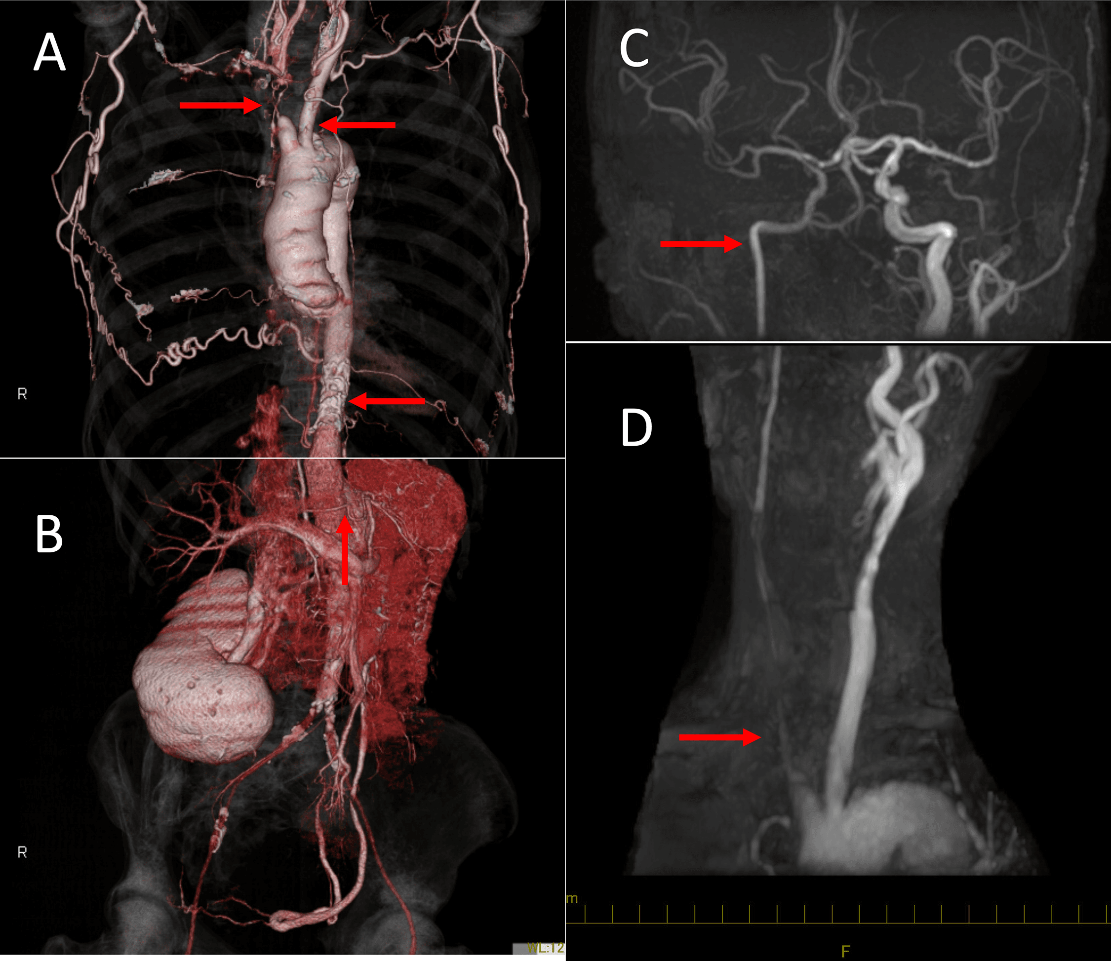
Figure 1. Preoperative imaging of arterial conditions. (A) Occlusion of the brachiocephalic artery, both subclavian arteries, and the origin of the celiac axis; (B) stenosis of the superior mesenteric artery; (C) stenosis of the left internal carotid artery; (D) occlusion of the right common carotid artery.
Surgical revascularization was planned. A partial median sternotomy was performed. The brachiocephalic artery was exposed and found to be narrowed at the occlusion site. The bilateral axillary arteries were accessed without nerve injury, and both the femoral arteries were exposed. Subcutaneous tunnels were created from the subclavian region to the femoral arteries. After systemic heparinization, the brachiocephalic artery was clamped, and a 14/7/7-mm J Graft SHIELD® (Japan Lifeline, Tokyo, Japan) was anastomosed. A 6-mm PROPATEN® graft (Gore, Tokyo, Japan) was then connected to the main body of the J Graft and extended to the right common carotid artery.
The right limb of the J Graft was tunneled to the subclavian region and connected to the right common femoral artery using an 8-mm PROPATEN® graft in an end-to-end fashion, followed by an end-to-side anastomosis to the artery. Another 6-mm PROPATEN® graft was used for a bypass from the right limb of the J Graft to the right axillary artery in an end-to-side fashion. This procedure was mirrored on the left side for both the axillary and common femoral arteries. Postoperative CTA confirmed graft patency (Fig. 2).
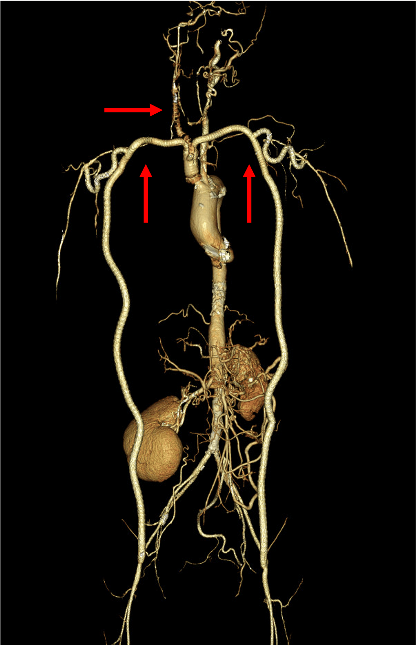
Figure 2. All bypass grafts were patent except for the right carotid artery graft.
To address progressive left carotid stenosis and prevent cerebral ischemia, a bypass from the brachiocephalic artery graft to the right common carotid artery was performed. Postoperatively, systolic blood pressure was maintained around 100 mmHg. On postoperative day 1, the patient developed headaches, but CT scans revealed no signs of cerebral hemorrhage. The headache persisted on day 2 despite continued blood pressure control. By postoperative day 5, a right-sided cerebral hemorrhage occurred, attributed to cerebral hyperperfusion syndrome (Figs. 3A, 3B). Catheterization at that time revealed significantly elevated ascending aortic pressure (280 mmHg), despite low peripheral readings, indicating uncontrolled systemic hypertension affecting cerebral circulation. To manage the hemorrhage, blood flow through the right common carotid artery bypass graft was surgically reduced by approximately 30% (Fig. 3C). The patient experienced improvement without any neurological sequelae following rehabilitation and expressed satisfaction with the resolution of limb ischemia. At the 1-year follow-up, rest pain had resolved immediately after revascularization, and the ABI values were 1.1 on the right and 1.0 on the left.
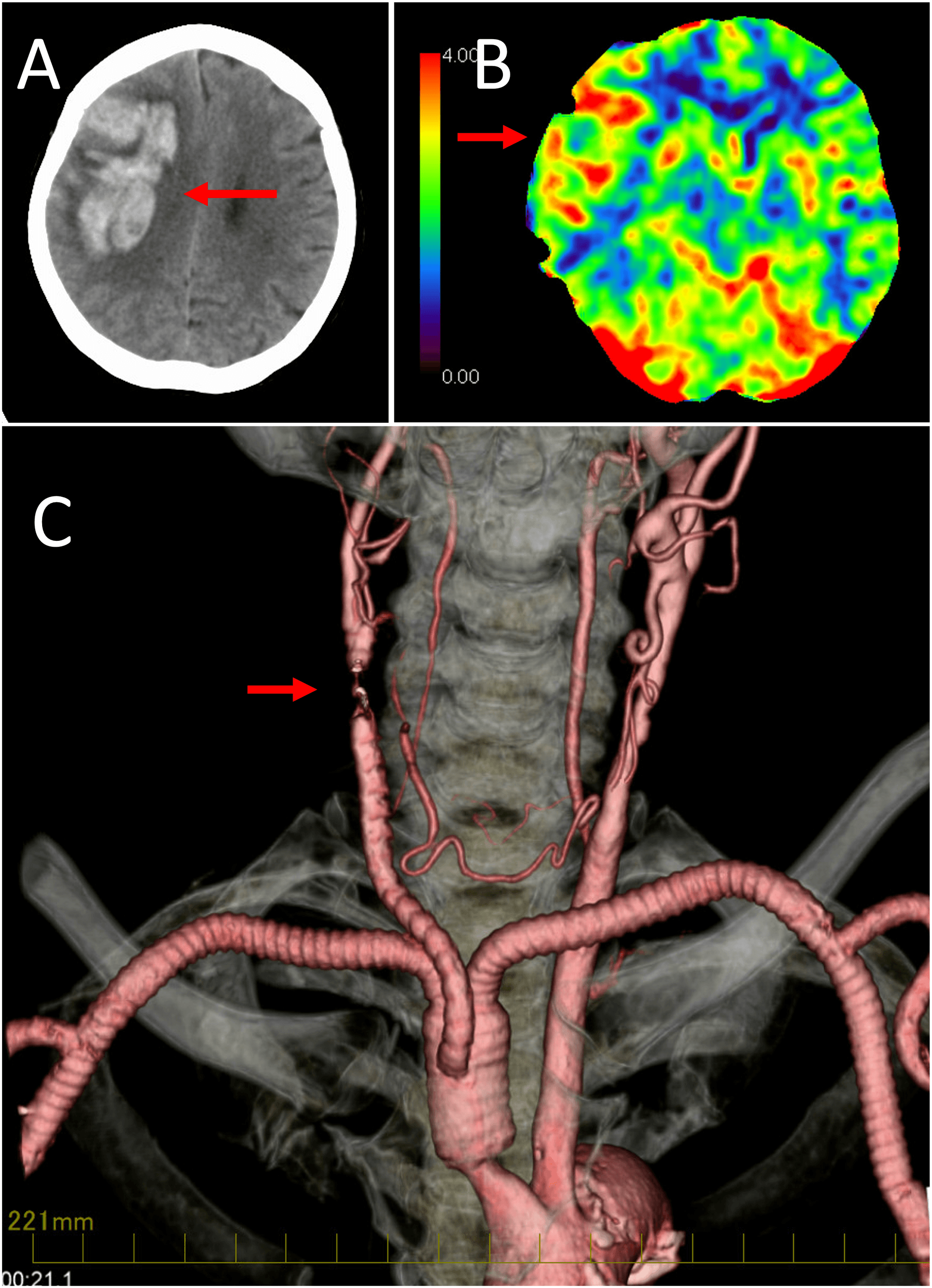
Figure 3. Changes in brain blood flow after bypass surgery. (A) Right cerebral hemorrhage; (B) hyperperfusion of the right brain after bypass surgery; (C) reduction of bypass graft flow.
Discussion
We encountered a case of Takayasu arteritis presenting with multivessel stenosis and occlusion, notably involving the brachiocephalic and carotid arteries. The patient underwent staged surgical revascularization aimed at preventing critical limb and cerebral ischemia. A key challenge was balancing timely revascularization with the risk of hyperperfusion injury, compounded by the difficulty of accurate blood pressure monitoring in a “pulseless” condition. Successful management strategies included utilizing proximal vessel stumps for bypass inflow, deliberately avoiding diseased arterial segments to reduce the risk of restenosis or pseudoaneurysm, and adapting perioperative care based on cerebral perfusion assessments. This case highlights the need for staged surgical planning, careful cerebral perfusion evaluation, and individualized revascularization strategies in Takayasu arteritis with complex vascular involvement.
Takayasu arteritis often manifests with aortic coarctation or aneurysm due to active aortic wall inflammation and may lead to renovascular hypertension requiring corticosteroid therapy prior to surgery.11 Clinical presentations frequently involve limb ischemia, cerebrovascular insufficiency, or renal impairment. Accurate blood pressure monitoring is often impeded by arterial occlusions, leading to the condition’s classification as “pulseless disease.” Compared with younger patients, in whom vascular lesions are often due to congenital or inflammatory causes, elderly patients more commonly present with calcified and fibrotic age-related changes that aggravate ischemia.12 Age at presentation also influences management, as older patients often benefit from less invasive endovascular approaches, while younger patients may be candidates for surgical reconstruction.13
In the stable phase, bypass grafting or segmental replacement is typically employed to relieve ischemia, while endovascular interventions are generally reserved for straightforward stenotic lesions.14,15 Surgical outcomes in middle aortic syndrome have generally demonstrated favorable long-term patency and blood pressure control, although perioperative risks remain significant.16 Reported series emphasize the importance of careful patient selection and individualized revascularization strategies.17 Our case aligns with these findings, highlighting both the technical feasibility and symptomatic improvement achievable with complex revascularization. In resource-limited settings, nonsurgical management with glucocorticoids and conventional immunosuppressants often remains the mainstay due to limited access to revascularization procedures.18 While effective in controlling inflammation, medical therapy alone is less successful in preventing ischemic complications, highlighting the disparity in outcomes compared to centers where surgical or endovascular options are available.
In this case, renal function was preserved, and no mesenteric ischemia was evident. Given the extensive occlusions, reconstruction of the right common carotid and limb arteries was pursued. Despite brachiocephalic artery occlusion, a 2-cm proximal stump allowed for safe debranching bypass inflow. Diseased segments were avoided to minimize suture-related complications and to ensure adequate graft length. A sternotomy-based debranching from the ascending aorta was considered, but it was deferred to preserve future surgical options for aortic root interventions. Endovascular treatment was evaluated as an alternative, but it posed considerable risks, including rupture, stent underexpansion, and restenosis due to active inflammation, as well as technical challenges presented by the complex arterial lesions.19
Strict systolic control was maintained for 2 weeks postoperatively to prevent rebleeding.20 This case underscores the critical importance of staged surgical planning in Takayasu arteritis with complex occlusions. Preoperative cerebral perfusion imaging is essential, even in asymptomatic carotid occlusions, to determine the necessity for and timing of carotid reconstruction.21 If collateral circulation is adequate, delaying carotid bypass may reduce the risk of hyperperfusion complications. Sequential cerebral perfusion assessments after limb revascularization can help guide further interventions safely.
Conclusion
In Takayasu arteritis with complex occlusions, staged revascularization guided by cerebral perfusion assessment is key to reducing complications such as hyperperfusion injury. Careful surgical planning, selective bypassing, and strict blood pressure control are essential for improving patient outcomes.
Acknowledgment
We thank the Department of Neurosurgery, Hirosaki University Hospital, for their intervention, and the patient’s collaboration in data collection.
Funding Source
No financial support was sought from any source.
Author Contributions
Z.Y. was responsible for data curation, data interpretation, and drafting of the original manuscript. K.M., Y.S., T.K., and M.M. made substantial contributions to revising the manuscript draft. All authors have read and approved the manuscript and agree with its content and data.
Data Availability
The datasets used in the current study can be obtained from the corresponding author upon reasonable request.
Ethical Statement
This article does not involve the participation of any animals. The patient provided written informed consent for the publication of this report and the accompanying images.
Conflict of Interest
The authors report no conflicts of interest in this work.
Supplemental Information
Supplemental information for this article can be found online at https://sup.jclinque.com/api/articles/82/download-suppl.
References
[1] Espitia O, Toquet C, Jamet B, Serfaty JM, Agard C. [Aortitis]. Rev Med Interne. December 2024;45(12):767–775. doi:10.1016/j.revmed.2024.06.015.
[2] Ahmed H, Ismayl M, Palicherla A, et al. A rare case of aortitis presenting as chest pain: a case report and literature review. Ann Med Surg (Lond). July 2024;86(7):4165–4169. doi:10.1097/ms9.0000000000002140.
[3] Benhuri B, ELJack A, Kahaleh B, Chakravarti R. Mechanism and biomarkers in aortitis–a review. J Mol Med (Berl). January 2020;98(1):11–23. doi:10.1007/s00109-019-01838-1.
[4] Johnston SL, Lock RJ, Gompels MM. Takayasu arteritis: a review. J Clin Pathol. July 2002;55(7):481–486. doi:10.1136/jcp.55.7.481.
[5] Shahriar Z, Hussen D, Reza S, Zahin A, Das A. Type V Takayasu arteritis with multivessel involvement: diagnostic challenges and imaging insights in a young female: a case report. Radiol Case Rep. August 2025;20(8):4032–4036. doi:10.1016/j.radcr.2025.04.118.
[6] Boparai N, Girgis M, Kiamanesh O, Al-Arnawoot A, Amad H, Tsang W. Takayasu arteritis causing aortitis and aortic regurgitation: a totally tubular case report. CASE (Phila). February 2021;5(1):62–66. doi:10.1016/j.case.2020.10.009.
[7] Takayasu M. A case with peculiar changes of the retinal central vessels. Acta Soc Ophthalmol Jpn. 1908;12:554–555.
[8] Gudbrandsson B, Molberg Ø, Garen T, Palm Ø. Prevalence, incidence, and disease characteristics of Takayasu arteritis by ethnic background: data from a large, population-based cohort resident in Southern Norway. Arthritis Care Res (Hoboken). February 2017;69(2):278–285. doi:10.1002/acr.22931.
[9] Gloor AD, Chollet L, Christ LA, Cullmann JL, Bonel HM, Villiger PM. Takayasu arteritis: prevalence and clinical presentation in Switzerland. PLoS One. 2021;16(6):e0250025. doi:10.1371/journal.pone.0250025.
[10] Watts RA, Hatemi G, Burns JC, Mohammad AJ. Global epidemiology of vasculitis. Nat Rev Rheumatol. January 2022;18(1):22–34. doi:10.1038/s41584-021-00718-8.
[11] Saadoun D, Bura-Riviere A, Comarmond C, Lambert M, Redheuil A, Mirault T. French recommendations for the management of Takayasu’s arteritis. Orphanet J Rare Dis. July 21, 2021;16(Suppl 3):311. doi:10.1186/s13023-021-01922-1.
[12] Yasuda T, Hagino N. A 50-year delay in the diagnosis of Takayasu arteritis: clinical presentation and therapeutic insights. Cureus. May 2025;17(5):e84361. doi:10.7759/cureus.84361.
[13] Oliveira JCS, Santos AMD, Aguiar MF, et al. Characteristics of older patients with Takayasu’s arteritis: a two-center, cross-sectional, retrospective Cohort study. Arq Bras Cardiol. 2023;120(1):e20220463. Particularidades dos Pacientes com Arterite de Takayasu em Idade Mais Avançada: Estudo Coorte, Retrospectivo e Transversal. doi:10.36660/abc.20220463.
[14] Adhikari B, Niraula B, Dahal P, Suvedi A, Subedi G. Challenges in diagnosis and management of Takayasu arteritis: a case report highlighting vascular complications and delayed recognition. Clin Case Rep. February 2025;13(2):e70174. doi:10.1002/ccr3.70174.
[15] Regola F, Uzzo M, Toniati P, Trezzi B, Sinico RA, Franceschini F. Novel therapies in Takayasu arteritis. Front Med (Lausanne). 2021;8:814075. doi:10.3389/fmed.2021.814075.
[16] Shin J, Cho A, Han A, Ahn S, Min S, Min SK. Long-term patency and complications of endovascular and surgical revascularization for Takayasu arteritis. Vasc Specialist Int. December 31, 2024;40:46. doi:10.5758/vsi.240090.
[17] Samaan M, Abramyan A, Sundararajan S, et al. Cerebrovascular implications of Takayasu arteritis: a review. Neuroradiology. January 2025;67(1):125–136. doi:10.1007/s00234-024-03472-2.
[18] Águeda AF, Monti S, Luqmani RA, et al. Management of Takayasu arteritis: a systematic literature review informing the 2018 update of the EULAR recommendation for the management of large vessel vasculitis. RMD Open. 2019;5(2):e001020. doi:10.1136/rmdopen-2019-001020.
[19] Naik G, Wadiwala I, Hiremath N. Endovascular treatment for aortic diseases: intercontinental disparities. Curr Cardiol Rep. July 21, 2025;27(1):115. doi:10.1007/s11886-025-02260-5.
[20] Koh NH, Kim SS, Oh HY, Kim S, Jang JW. Hyperperfusion syndrome following tissue plasminogen activator administration: a case report with radiological evidence. J Korean Soc Radiol. November 2024;85(6):1200–1208. doi:10.3348/jksr.2024.0023.
[21] Inoue Y, Inoue M, Koga M, Matsuda H. Preoperative brain computed tomographic perfusion for quantitative evaluation of cerebral malperfusion caused by acute type A aortic dissection. JTCVS Tech. December 2021;10:190–195. doi:10.1016/j.xjtc.2021.09.031.
 |
Copyright: © 2025 Yu et al. This work is licensed under a Creative Commons Attribution 4.0 International License, which permits unrestricted use, distribution, and reproduction in any medium, provided the original work is properly cited. |