| Journal of Clinical Question. 2025; 2(2): e63 https://doi.org/10.69854/jcq.2024.0020 Advance access publication date 27 March 2025 |
 |
Case Report
Surgical Site Infection Caused by Serratia marcescens Following Adult Spinal Deformity Surgery: A Case Report
1Department of Spine Surgery, Shinagawa Shisyokai Hospital, Tokyo, Japan.
2Department of Neurosurgery, Shinagawa Shisyokai Hospital, Tokyo, Japan.
3Department of Spine Surgery, Heiwakai Heiwa Hospital, Yokohama, Japan.
*Corresponding Author: Email: youngcmu@gmail.com
Submitted: November 29, 2024 Accepted: March 27, 2025
Clinical Question Box
What are the optimal strategies for managing a Serratia marcescens surgical site infection following adult spinal deformity surgery?
The management of deep surgical site infections (SSIs) following adult spinal deformity surgery primarily involves thorough debridement, intravenous antibiotic administration, and implant removal. If these measures are insufficient, antibiotic-impregnated calcium phosphate bone cement paste can be used for targeted local therapy, offering a synergistic antibacterial effect. This approach is particularly beneficial for early-onset deep SSIs in cases where implant retention is necessary due to the high risk of spinal instability.
Abstract
Background: Surgical site infections (SSIs) are severe complications of spinal surgeries. Serratia marcescens, an increasingly recognized pathogen causing nosocomial infections, presents unique challenges in managing SSIs, particularly in spinal instrumentation cases. Case Presentation: A 68-year-old female with a history of osteoporosis developed a Serratia marcescens infection following staged adult spinal deformity (ASD) surgery. The infection persisted despite seven aggressive debridement procedures and systemic antibiotic therapies. During the final debridement, antibiotic-impregnated calcium phosphate bone cement paste (CPC) was applied to the surgical site to augment all the pedicle screws and infectious disc spaces. This approach helped successfully manage the infection without resorting to spinal implant removal. The patient has remained symptom-free for 5 years since this treatment. Conclusion: SSIs caused by Serratia marcescens present a significant challenge in ASD surgery. Antibiotic-impregnated CPC enabled sustained local antibiotic release, maintaining a high concentration in the long term and contributing to infection resolution without requiring implant removal.
Keywords: Serratia marcescens, surgical site infection, calcium phosphate bone cement, adult spinal deformity surgery
Introduction
Surgical site infections (SSIs) are significant complications in spinal surgeries, which can often lead to prolonged recovery times and challenging management.1 Among the pathogens that cause SSIs, Serratia marcescens, a Gram-negative, rod-shaped bacterium, has been recognized as an emerging cause of nosocomial infections.2 Recent studies indicate the rising prevalence of Serratia marcescens infections in patients after surgery and their potential for antimicrobial resistance, emphasizing the clinical importance of addressing the infections caused by this pathogen.3,4
SSIs caused by Serratia marcescens pose unique challenges, particularly in cases requiring extensive instrumentation, prolonged operative times, and significant blood loss, as seen in adult spinal deformity (ASD) surgeries.5 These factors and the pathogen’s resistance profile complicate diagnosis and treatment.6 This case report describes a rare case of SSI caused by Serratia marcescens following an ASD surgery, highlighting the clinical course of the infection and evaluating the effectiveness of antibiotic-impregnated calcium phosphate bone cement paste (CPC) in controlling the infection without requiring implant removal. This approach underscores the potential of local antibiotic delivery in achieving long-term infection resolution in similar cases.
Case Presentation
A 68-year-old female with a history of an osteoporotic T12 fracture presented with progressive kyphoscoliosis, low back pain, trunk stiffness, and worsening fatigability over 5 years. She also experienced horizontal gaze disturbance due to global sagittal malalignment. She was referred to our clinic for surgical intervention. The complexity of her spinal deformity (Fig. 1A) necessitated staged posterior surgeries.
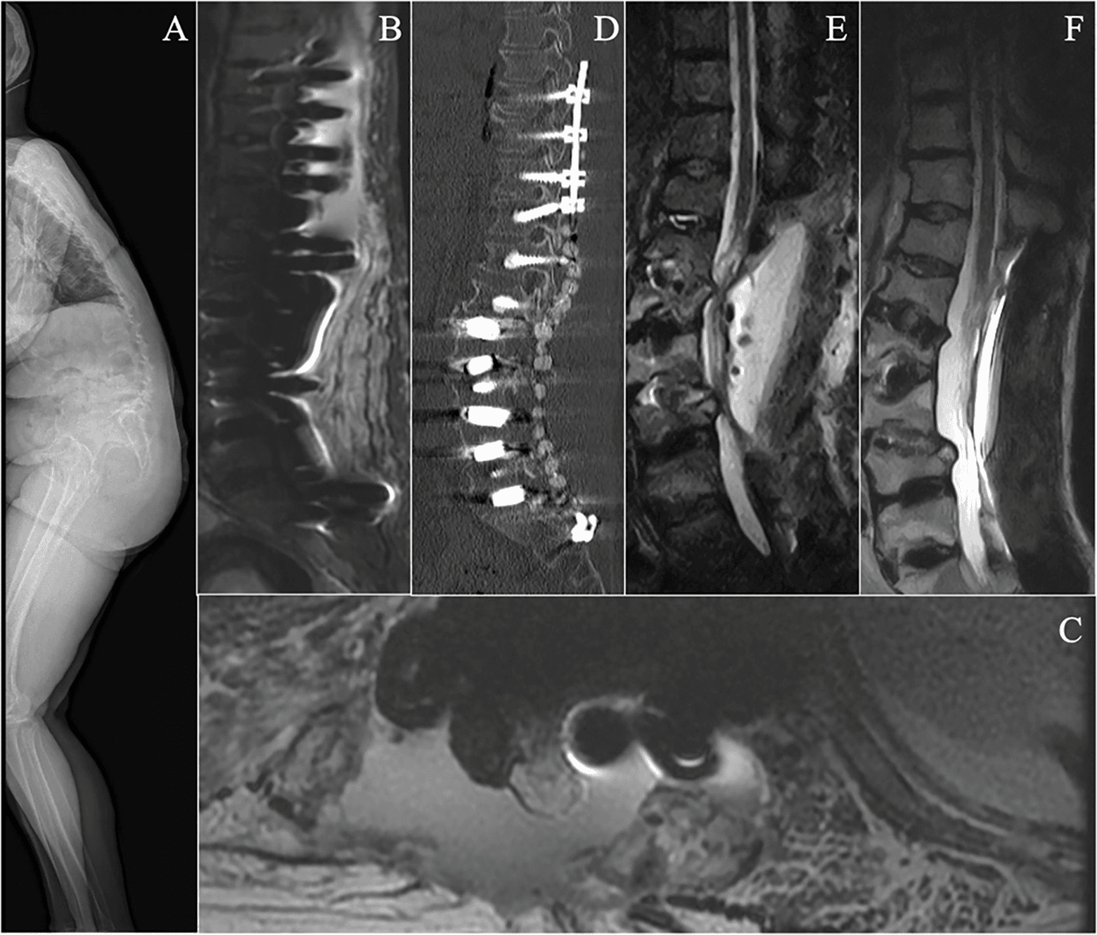
Figure 1. Imaging findings of the patient. (A) Preoperative standing full-body radiograph; (B and C) STIR sequence and T2-weighted MRI taken the day after the second debridement surgery; (D) sagittal CT showing cement beads in place; (E) MRI STIR sequence showing fluid signal at the surgical site before the final debridement surgery; and (F) T2-weighted MRI showing improvement at the infection site before discharge.
In the first stage, pedicle screws were inserted from T8 to the sacroiliac joints via a posterior midline incision. On postoperative day 9 (POD 9), no complications were observed. L2 and L4 pedicle subtraction osteotomies (PSOs) and multilevel lumbar interbody fusions were performed in the second stage. On POD 12, 2 days after the second-stage surgery, the patient’s maximum body temperature rose to 38.8°C. On POD 13, the C-reactive protein (CRP) level reached 27.4 mg/dL. These findings were initially considered a typical postoperative inflammatory response due to the invasive nature of the surgery. Prophylactic cefazolin was administered to prevent SSI (Fig. 2).
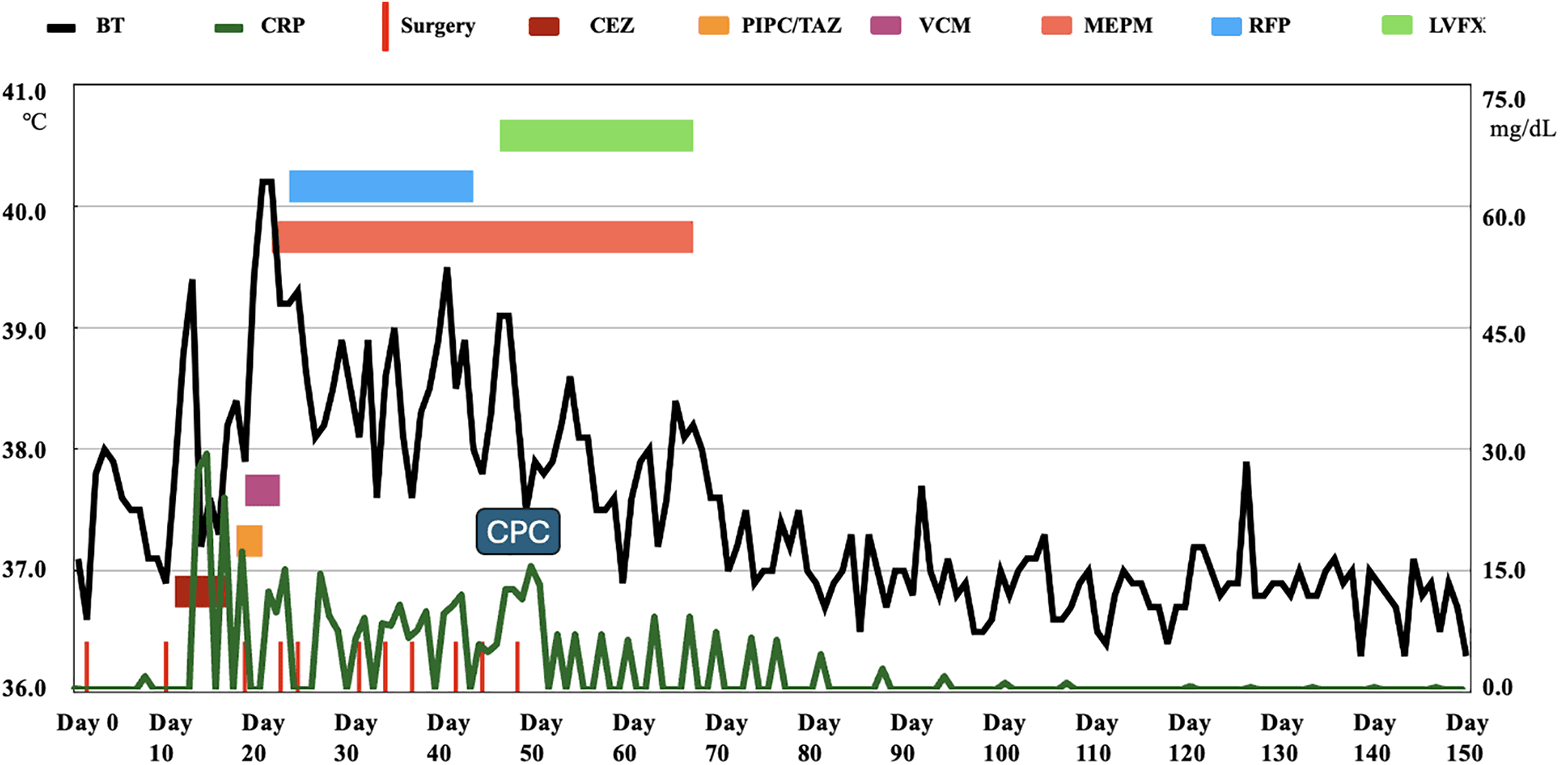
Figure 2. Clinical Course of the Patient. BT: body temperature, CRP: C-reactive protein, CEZ: cefazolin, PIPC/TAZ: piperacillin/tazobactam, VCM: vancomycin, MEPM: meropenem, RFP: rifampicin, LVFX: levofloxacin, CPC: calcium phosphate bone cement.
On POD 15, wound dehiscence with discharge was observed, raising a suspicion of SSI, which was confirmed through magnetic resonance imaging (MRI) (Figs. 1B, 1C). Blood culture and wound secretion samples were obtained. Initial prophylactic cefazolin was escalated to piperacillin/tazobactam and subsequently to vancomycin (VCM) to target Staphylococcus spp.7 On POD 18, the first debridement surgery was performed. Microbiological cultures revealed the presence of Serratia marcescens in both the wound secretion and blood samples. Based on antibiotic susceptibility testing, intravenous antibiotics were switched to meropenem (MEPM) on POD 21. A second debridement was performed on POD 25. The patient’s condition deteriorated acutely on POD 26; she presented with hypoproteinemia and pleural effusion, necessitating transfer to a general hospital. During the 23-day hospitalization that followed, the patient underwent five additional debridement surgeries and received intravenous MEPM and oral rifampin.
Despite a total of seven aggressive surgical debridements, including pedicle screw removal, soaking in povidone-iodine, and reinsertion, the infection persisted. Then, VCM-loaded cement beads were placed beneath the muscles (Fig. 1D). However, the patient continued to experience wound discharge, erythema, swelling, fever, elevated CRP levels, and persistent abnormalities that were revealed on the MRI (Fig. 1E). Intravenous levofloxacin (LVFX) was initiated on POD 47.
The final debridement was performed on POD 49, as all previous treatments had failed. Antibiotic-impregnated CPC (MEPM: 3.5 g with 84 g CPC; minocycline (MINO): 3 g with 72 g CPC; antibiotic ratio: 4%) was used to augment all pedicle screws and serve as a local antibiotic delivery system. The CPC paste was shaped into thin rods and inserted into the patient’s vertebral bodies via a 4.2-mm-diameter inserter, followed by pedicle screw reinsertion within 10 minutes to avoid the risk of CPC solidification. CPC was also placed into the L2/3, L3/4, L4/5, and L5/S1 disc spaces, where discitis was evident on the MRI (Fig. 1E). Following the final surgery, a Clostridioides difficile toxin test revealed negative despite the patient experiencing diarrhea. Intravenous MEPM and LVFX were continued for 20 days until fever and CRP normalized, after which the regimen was switched to oral LVFX.
After a 5-month hospital stay, a follow-up MRI scan showed improvement at the infection site (Fig. 1F). The patient was then discharged with a prescription for a 3-month course of oral LVFX. At the 5-year follow-up, there was no recurrence of infection, the patient’s preoperative symptoms had all resolved, and no long-term complications related to the ASD surgery were observed.
Discussion
Prolonged operation time and increased blood loss are significant risk factors that can lead to postoperative complications in ASD surgery.8 Staged surgery can mitigate perioperative complications in high-risk cases.9
The incidence of SSI in spinal instrumentation surgery is approximately 1.8%, with Staphylococcus aureus being the most common pathogen.1,5 To our knowledge, this is the first reported case of SSI caused by Serratia marcescens following ASD surgery. Given the high mortality rate related to Serratia marcescens infections (31%),10 early and accurate diagnosis is crucial. While microbiological cultures remain the gold standard for diagnosing SSI, clinical indicators such as wound characteristics (redness, swelling, secretions, and dehiscence), high fever, elevated CRP levels, and imaging findings can also provide essential diagnostic clues.11
A previous study reported that in 42.9% of SSI cases, elevated CRP levels were observed on PODs 7 and 8, followed by a second CRP peak on POD 12 ± 2.3. A CRP level of 22.5 mg/dL was identified as the cut-off value for detecting infection (sensitivity: 92.9%; specificity: 78.2%).12 In the current case, elevated CRP levels were recorded on day 12 after the first-stage surgery, suggesting that the infection may have developed following this initial procedure. We monitored the CRP level every 1–3 days during the early stages of treatment. The elevated CRP levels correlated with the patient’s worsening general condition. Although metal artifacts in MRI can be a concern in such cases, short tau or short TI inversion recovery (STIR) sequences are more effective than T1- and T2-weighted imaging for detecting spinal osteomyelitis.13 Postoperative MRI confirmed a massive subcutaneous fluid collection and bone edema in this case, indicating ongoing infection at the surgical site. The final MRI before discharge showed a remarkable reduction in subcutaneous fluid. The source of the Serratia marcescens infection remains unclear but may be associated with poor oral hygiene or the first surgical intervention.
Management of spinal infections involving instrumentation is challenging, as 33% of cases require implant removal.5,14 Early infections can often be managed with antibiotics alone (62.5%),15 while late infections typically necessitate implant removal due to biofilm formation. In the current case, removing the implants posed a high risk of spinal instability, given the structural compromise posed by the L2 and L4 PSO. The infection persisted despite repeated debridement and the administration of intravenous antibiotics, possibly due to inadequate elimination of the pathogen and/or insufficient local concentration of intravenously administered antibiotics.
Antibiotic-impregnated CPC is a practical approach for delivering local antibiotic therapy to treat established orthopedic infections.16 CPC+VCM could release 5.5 times more VCM than polymethylmethacrylate (PMMA)+VCM and sustain release for over 8 weeks, as compared to VCM release from PMMA+VCM, which ceases after 4 weeks in vivo.17 The compression strength of CPC is related to the type and amount of antibiotics used. Increasing the antibiotic concentration reduces the compression strength. The compression strength of 5% CPC+VCM is about two-thirds that of CPC.18 In this case, we used 4% antibiotic-impregnated CPC, which is easy to mix in practice and balances compression strength with antimicrobial efficacy. Based on our experiences, we propose that in cases where the pathogen is unknown, a mixture of CPC+VCM and CPC+MEPM could be used for empirical treatment. However, further investigation is needed in this regard.
Antibiotic resistance poses a significant challenge in treating Serratia marcescens infections. As no standard antibiotic regimen exists for this infection, the results of microbiological cultures are crucial for guiding therapy. In this case, in addition to two urine cultures, we performed seven wound secretion swabs, four blood cultures, and one tip-of-drain culture at different stages of treatment. The first three blood cultures, three wound secretion cultures, and the tip-of-drain culture tested positive for Serratia marcescens, confirming the presence of the pathogen and underscoring the need for targeted antibiotic therapy based on antimicrobial susceptibility testing. Although colistin, tigecycline, and carbapenem are the current first-line choices,10 increasing resistance to carbapenem has been observed.2 In the present case, intravenous MEPM was administered for 27 days before the final debridement surgery, yet persistent high fever and elevated CRP suggested possible resistance to MEPM. MEPM- and MINO-impregnated CPC, combined with intravenous antibiotics, demonstrated a synergistic effect. As a result, the patient’s fever and CRP levels normalized, highlighting the success of this combined therapeutic strategy. Long-term oral antibiotic treatment lacks robust evidence and clear guidelines for this pathogen. Treatment duration should be individualized based on clinical response, infection severity, and risk–benefit considerations.
Conclusion
This case highlights the challenges of managing Serratia marcescens infections after ASD surgery, where antibiotic resistance and limited local antibiotic concentration complicate treatment. Despite these challenges, 4% antibiotic-impregnated CPC enabled sustained local antibiotic release, maintaining a high antibiotic concentration in the long term and contributing to infection resolution without requiring implant removal.
Acknowledgment
We appreciate the patient’s cooperation in data collection.
Funding Source
No financial support was sought from any source.
Author Contributions
B.Y. was responsible for data curation, data interpretation, and drafting of the original manuscript. T.M. and K.O. made substantial contributions to revising the manuscript draft. All authors have read and approved the manuscript and agree with its content and data.
Data Availability
The datasets used in the current study can be obtained from the corresponding author upon reasonable request.
Ethical Statement
This article does not involve the participation of any animals. The patient provided written informed consent for the publication of this report and accompanying images.
Conflict of Interest
The authors report no conflicts of interest in this work.
Supplemental Information
Supplemental information for this article can be found online at https://sup.jclinque.com/api/articles/63/download-suppl.
References
[1]. Zhang T, Lian X, Chen Y, Cai B, Xu J. Clinical outcome of postoperative surgical site infections in patients with posterior thoracolumbar and lumbar instrumentation. J Hosp Infect. 2022;128(Suppl):26–35. doi:10.1016/j.jhin.2022.06.014.
[2]. Cosimato I, Santella B, Rufolo S, et al. Current epidemiological status and antibiotic resistance profile of serratia marcescens. Antibiotics. 2024;13(4):323. doi:10.3390/antibiotics13040323.
[3]. Tavares-Carreon F, De Anda-Mora K, Rojas-Barrera IC, Andrade A. Serratia marcescens antibiotic resistance mechanisms of an opportunistic pathogen: a literature review. PeerJ. 2023;11:e14399. doi:10.7717/peerj.14399.
[4]. Lynch JP, Clark NM, Zhanel GG. Escalating antimicrobial resistance among Enterobacteriaceae: focus on carbapenemases. Expert Opin Pharmacother. 2021;22(11):1455–1473. doi:10.1080/14656566.2021.1904891.
[5]. Konishi K, Sano H, Kawano Y, et al. Factors related to surgical site infection in spinal instrumentation surgery: a retrospective study in Japan. Asian Spine J. Published Online. October 22, 2024;22(6):822–828. doi:10.31616/asj.2024.0274.
[6]. Mancuso G, Midiri A, Gerace E, Biondo C. Bacterial antibiotic resistance: the most critical pathogens. Pathog Basel Switz. 2021;10(10):1310. doi:10.3390/pathogens10101310.
[7]. Chen SH, Lee CH, Huang KC, Hsieh PH, Tsai SY. Postoperative wound infection after posterior spinal instrumentation: analysis of long-term treatment outcomes. Eur Spine J. 2015;24(3):561–570. doi:10.1007/s00586-014-3636-9.
[8]. Lee NJ, Kothari P, Kim JS, et al. Early complications and outcomes in adult spinal deformity surgery: an NSQIP study based on 5803 patients. Glob Spine J. 2017;7(5):432–440. doi:10.1177/2192568217699384.
[9]. Rhee JM, Bridwell KH, Lenke LG, et al. Staged posterior surgery for severe adult spinal deformity. Spine. 2003;28(18):2116–2121. doi:10.1097/01.BRS.0000090890.02906.A4.
[10]. Zivkovic Zaric R, Zaric M, Sekulic M, et al. Antimicrobial treatment of serratia marcescens invasive infections. Antibiotics. 2023;12(2):367. doi:10.3390/antibiotics12020367.
[11]. Lim S, Yoo YM, Kim KH. No more tears from surgical site infections in interventional pain management. Korean J Pain. 2023;36(1):11–50. doi:10.3344/kjp.22397.
[12]. Hoeller S, Roch PJ, Weiser L, Hubert J, Lehmann W, Saul D. C-reactive protein in spinal surgery: more predictive than prehistoric. Eur Spine J Off Publ Eur Spine Soc Eur Spinal Deform Soc Eur Sect Cerv Spine Res Soc. 2021;30(5):1261–1269. doi:10.1007/s00586-021-06782-8.
[13]. Feuerriegel GC, Sutter R. Managing hardware-related metal artifacts in MRI: current and evolving techniques. Skeletal Radiol. 2024;53(9):1737–1750. doi:10.1007/s00256-024-04624-4.
[14]. Hersh A, Young R, Pennington Z, et al. Removal of instrumentation for postoperative spine infection: systematic review. Published Online. July 9, 2021;35(3):376–388. doi:10.3171/2020.12.SPINE201300.
[15]. Dobran M, Marini A, Gladi M, et al. Deep spinal infection in instrumented spinal surgery: diagnostic factors and therapy. Il G Chir. 2017;38(3):124–129. doi:10.11138/gchir/2017.38.3.124.
[16]. Niikura T, Lee SY, Iwakura T, Sakai Y, Kuroda R, Kurosaka M. Antibiotic-impregnated calcium phosphate cement as part of a comprehensive treatment for patients with established orthopaedic infection. J Orthop Sci Off J Jpn Orthop Assoc. 2016;21(4):539–545. doi:10.1016/j.jos.2016.05.003.
[17]. Mukai M, Uchida K, Sugo K, et al. Long-term antibacterial activity of vancomycin from calcium phosphate cement in vivo. Biomed Mater Eng. 2022;33(1):41–50. doi:10.3233/BME-211243.
[18]. Suzuki M, Tsukeoka T, Tsuneizumi Y, et al. Mechanical strength and in vitro antibiotic release profile of antibiotics-loaded calcium phosphate bone paste. Clin Orthop Surg. 2004;39(3):309–314.
 |
Copyright: © 2025 Yang et al. This work is licensed under a Creative Commons Attribution 4.0 International License, which permits unrestricted use, distribution, and reproduction in any medium, provided the original work is properly cited. |