| Journal of Clinical Question. 2025; 2(6): e100 https://doi.org/10.69854/jcq.2025.0039 Advance access publication date 30 December 2025 |
 |
Review
RSV-Induced Epigenetic Reprogramming: Mechanisms and Therapeutic Opportunities
Guangyuan Yu1,2,3,#, Mohan Wang4,#, Heqian Fan4,#, Yuanyuan Li4, Sisi Chen5, Jinghe Li6,*, Zi Lv 7,8,9,*
1Department of Gastroenterology, Pediatric Research Institute, Children’s Hospital of Chongqing Medical University, Chongqing, China.
2National Clinical Research Center for Child Health and Disorders, Ministry of Education Key Laboratory of Child Development and Disorders, Chongqing, China.
3Chongqing Key Laboratory of Child Rare Diseases in Infection and Immunity, Chongqing, China.
4Chongqing Medical University, Chongqing, China.
5Chongqing Medical and Pharmaceutical College, Chongqing, China.
6Institute of Reproductive Health and Perinatology, Guangzhou Women and Children’s Medical Center, Guangzhou Medical University, Guangzhou, China.
7Reproductive Medicine Center, The First Affiliated Hospital of Chongqing Medical University, Chongqing, China.
8Joint International Research Laboratory of Reproduction & Development, Ministry of Education, The First Affiliated Hospital of Chongqing Medical University, Chongqing, China.
9The Innovation and Talent Recruitment Base of Maternal-Fetal Medicine, The First Affiliated Hospital of Chongqing Medical University, Chongqing, China.
#All authors share first authorship
*Corresponding Authors: Jinghe Li: jinghe_li_0514@yahoo.co.jp; Zi Lv: lvzi0121@foxmail.com
Submitted: November 02, 2025 Accepted: December 29, 2025
Clinical Question Box
Can epigenetic biomarkers help determine which infants with respiratory syncytial virus (RSV) infection are at greater risk for severe disease?
RSV infection causes specific epigenetic changes, including variations in DNA methylation, histone modifications, and shifts in noncoding RNA levels. These molecular markers reflect the strength of the antiviral and inflammatory responses and are linked to both immediate disease severity and future respiratory issues. Detecting these epigenetic signals early will enable healthcare providers to identify infants at higher risk of deterioration or long-term respiratory problems, such as recurrent wheeze or asthma. Incorporating epigenetic biomarkers into clinical evaluation could allow earlier risk assessment and more tailored treatment plans compared to symptom-based approaches.
Abstract
Respiratory syncytial virus (RSV) is a leading cause of severe lower respiratory infections in infants and young children, and it significantly contributes to illness in older adults and immunocompromised individuals. Besides established immune evasion mechanisms, growing evidence indicates that RSV actively reprograms host epigenetic regulation, affecting both antiviral defenses and long-term respiratory health. RSV alters DNA methylation, histone modifications, chromatin accessibility, and noncoding RNA (ncRNA) expression in various immune and structural cell types, including airway epithelial cells, T cells, B cells, and innate immune cells. These epigenetic changes influence transcriptional programs that regulate cytokine responses, cytotoxic activity, and memory development, ultimately impacting disease severity and the risk of chronic issues like recurrent wheeze and asthma. New epigenetic biomarkers offer promising options for early risk assessment and personalized prognosis in clinical settings. Epigenetic-targeted treatments, such as selective histone deacetylase inhibitors and CRISPR (Clustered Regularly Interspaced Short Palindromic Repeats)-based gene regulation, show therapeutic promise, though challenges remain regarding off-target effects, delivery methods, and cell-specific targeting. Advances in multi-omics integration, spatial and single-cell technologies, and patient-derived airway organoids provide increasingly relevant human models for studying RSV–epigenome interactions and developing precise therapies. Understanding and harnessing epigenetic reprogramming could spawn novel approaches for predicting severe disease and preventing long-term complications.
Keywords: Respiratory syncytial virus, epigenetics, immune evasion, biomarkers, therapeutics
Introduction
Respiratory syncytial virus (RSV) is a single-stranded, negative-sense RNA virus belonging to the Pneumoviridae family. The RSV genome encodes structural and nonstructural proteins that collectively govern viral entry, replication, and immune evasion.1 The attachment glycoprotein facilitates binding to host cells through receptors such as heparan sulfate proteoglycan and fractalkine receptor (C-X3-C motif chemokine receptor 1 [(CX3CR1)]), enabling fusion and entry mediated by the fusion glycoprotein (F).2–4 The small hydrophobic protein modulates host immunity,5 while the matrix protein coordinates viral assembly.6 The nucleoprotein, phosphoprotein, and polymerase complex carry out genomic replication and transcription.7,8 Nonstructural proteins 1 (NS1) and 2 (NS2) are key antagonists of the host innate immune response. Through these proteins, RSV disrupts multiple innate immune pathways, including toll-like receptor and retinoic acid–inducible gene I (RIG-I)-like receptor signaling and inflammasome activation.2,9–16 NS1 and NS2 inhibit type I interferon production by targeting interferon regulatory factors (IRFs) and signal transducer and activator of transcription 2; they also shift the cytokine response toward an immunosuppressive profile. Similarly, the F protein can inhibit interferon (IFN) production and leukocyte recruitment.3,17,18 These viral strategies foster a T helper type 2 (Th2)–biased immune environment, resulting in long-term effects that elevate the risk of childhood asthma.19,20 While these classical mechanisms explain much of RSV’s effective immune evasion, emerging evidence indicates that the virus also co-opts host epigenetic mechanisms to facilitate persistent replication and immune modulation.
In 2019 alone, RSV caused an estimated 33 million episodes of acute lower respiratory tract infections in young children, leading to 3.6 million hospitalizations.21 It is a leading global cause of these infections in children under 5. Nearly all children are infected at least once by age 2,22 and reinfection remains common throughout life because natural immunity does not provide full protection. Severe cases also occur in the elderly and immunocompromised individuals.23–25 RSV follows a distinct seasonal pattern. In temperate climates of the Northern Hemisphere, outbreaks usually occur from October or November through April or May, peaking in January or February. In the Southern Hemisphere, peaks occur from May to September.26 In tropical regions, RSV circulation is often linked to rainy seasons.27 Pandemic-related changes in social behavior altered these patterns, with intense off-season surges documented when coronavirus disease 2019 (COVID-19) mitigation measures were lifted.28 Transmission occurs mainly through inoculation of the nasopharynx or eyes after direct contact with virus-containing secretions or contaminated surfaces. RSV is highly durable in the environment; it can survive for several hours on hands and fomites, facilitating quick spread in households and healthcare settings. Studies show that 42% of RSV infections may be asymptomatic, but affected individuals still shed the virus for an average of 11 days, posing an ongoing transmission risk.29
RSV disease severity varies considerably among individuals and is influenced by host-related factors. Established risk factors include prematurity, chronic lung disease, congenital heart disease, Down syndrome, and immunocompromising conditions.30 Social and environmental factors also contribute: infants who attend daycare or live with older siblings are more likely to acquire infection, and exposure to secondhand smoke increases severity.31 Genetic studies further suggest that polymorphisms in cytokine- and immune-regulation genes, including interleukin-4 (IL-4), IL-8, IL-10, IL-13, surfactant proteins A and D, and toll-like receptor genes, are associated with greater susceptibility to severe RSV.32 Collectively, these epidemiological data demonstrate that RSV is not only ubiquitous but maintains a strong seasonal presence and imposes a disproportionate burden on vulnerable individuals.
Clinical Features
RSV in Adults
Although RSV has traditionally been labeled a “pediatric virus,” it is increasingly recognized as a significant pathogen in adults, especially older adults and immunocompromised patients. While healthy adults usually experience a mild upper respiratory infection, outcomes are much worse in high-risk groups. In adults undergoing hematopoietic cell transplantation or with hematologic cancers, progression to a lower respiratory tract infection is common and associated with high mortality.33 An observational study indicates that among severely immunocompromised adults with RSV who require intensive care unit (ICU) admission, approximately 50% require mechanical ventilation.34 In these populations, RSV can cause respiratory failure, longer hospital stays, and death.
Treatment in adults remains limited and is primarily supportive, though pharmacological therapy is considered in high-risk situations. Ribavirin, a nucleoside analog with in vitro antiviral activity against RSV, is approved by the Food and Drug Administration (FDA) and can be administered orally or via aerosol.35 Clinical practice often combines ribavirin with intravenous immune globulin and, occasionally, glucocorticoids, based on observational studies indicating reduced progression from upper to lower respiratory tract infections and lower mortality. However, ribavirin use is restricted by toxicities such as hemolytic anemia, bronchospasm, and teratogenic effects during pregnancy. RSV is also clinically significant in adults with asthma or chronic obstructive pulmonary disease, as infection can trigger exacerbations and worsen underlying conditions. Therefore, although pediatric cases dominate RSV-related statistics, the disease also contributes substantially to adult respiratory morbidity, especially in frail or medically complex populations.
RSV in Infants and Children
RSV causes the highest disease burden in infants and young children and is recognized as the leading cause of hospitalization for lower respiratory tract infections, such as bronchiolitis and pneumonia, in infants aged less than 1 year. Globally, RSV leads to hospitalization in 0.4% of children under 5 each year, with the highest burden among infants under 6 months, reaching hospitalization rates of 2%–6%, especially in preterm infants.36 In an international multicenter case–control study, RSV accounted for 31% of severe pneumonia hospitalizations among children aged 1–59 months in Africa and Asia.37 A prospective European birth cohort showed that 2% of healthy full-term infants are hospitalized during their first RSV season.38
RSV is also a common cause of outpatient visits. Prospective, population-based surveillance showed that RSV accounted for roughly 10%–15% of medically attended acute respiratory infections in outpatient and emergency department settings among young children, with the highest incidence occurring in early infancy.39 Mortality rates vary significantly across settings. In high-resource countries, RSV-associated mortality in children under 5 is very low (well below 1 death per 100,000 per year).21 Conversely, in low-resource regions, RSV is a major cause of childhood mortality, accounting for up to 6.5% of deaths among infants aged 28 days to <6 months,40 ranking second only to malaria. Importantly, approximately 20% of fatal pediatric RSV cases occur in children without major underlying risk factors,41 and nosocomial infection accounts for about 20% of reported in-hospital RSV deaths globally (up to ~26% in high-income settings).30 Due to its high transmissibility and substantial clinical burden in early infancy, RSV remains a primary target for preventive therapies, especially for neonates and high-risk infants.
Treatment and Prevention Strategies
Management of RSV infection in both adults and children is mainly supportive, focusing on maintaining hydration, oxygenation, and proper ventilation. Supportive care includes regular monitoring, suctioning of secretions, hydration, and escalation to supplemental oxygen or mechanical ventilation when necessary. Among hospitalized infants with RSV, only a small minority require invasive mechanical ventilation (about 2% in a prospective multicountry cohort),42 whereas among severely immunocompromised adults admitted to the ICU for RSV, approximately half require mechanical ventilation. Antiviral treatments are limited. Ribavirin may be considered for severely ill or immunocompromised pediatric patients, but is not routinely recommended for otherwise healthy infants due to unclear benefit and potential side effects. Intravenous immunoglobulin with high RSV-neutralizing activity has been shown to reduce viral load and prevent disease in animal studies, but is no longer commercially available.
A significant change in RSV prevention is underway. Both Arexvy and Abrysvo are authorized by the FDA for RSV prevention, but only Abrysvo is approved for use in pregnant individuals.43 Evidence indicates that infants with higher RSV antibody levels have milder disease and are less likely to develop lower respiratory tract infections. Two preventive strategies are now available: maternal RSV vaccination in the third trimester, which enables passive transfer of RSV antibodies to the newborn, and long-acting monoclonal antibody prophylaxis (nirsevimab), administered either before RSV season or at birth to protect infants throughout the season.44 Both methods significantly decrease RSV-related hospitalizations, representing the most essential progress in RSV prevention in decades. A recent study showed the efficacy of clesrovimab in preventing RSV disease in healthy infants.45
Epigenetic Mechanisms in RSV Infection
Epigenetics involves the study of heritable changes in gene expression that occur without alterations to the underlying DNA sequence. Key epigenetic mechanisms include DNA methylation, histone modifications, ncRNAs, and chromatin remodeling.46–48 DNA methylation entails the covalent addition of a methyl group to cytosine bases in cytosine–phosphate–guanine (CpG) dinucleotides, typically leading to transcriptional silencing by obstructing transcription factor binding.49 Histone modifications are post-translational changes to histone tails, such as acetylation, methylation, phosphorylation, ubiquitination, and the recently identified lactylation, that dynamically regulate chromatin structure and gene transcription.47,48,50–52 ncRNAs, including microRNAs (miRNAs) and long ncRNAs (lncRNAs), fine-tune gene expression post-transcriptionally; for instance, miRNAs can inhibit translation or promote messenger RNA (mRNA) degradation by binding to target transcripts.53 Chromatin remodeling involves ATP-dependent complexes that alter nucleosome positioning to regulate DNA accessibility and transcription.54 RSV infection disrupts these regulatory layers by interacting with host methyltransferases, histone modifiers, and ncRNA pathways, leading to altered DNA methylation patterns, aberrant histone marks, and dysregulated ncRNA expression. These changes collectively shape antiviral gene networks and viral replication.50,52 Understanding how RSV exploits host epigenetic machinery may reveal novel antiviral targets.
Epithelial Cell Epigenetic Remodeling and Its Barrier Defense
As the primary target of RSV and the first line of defense against infection, respiratory epithelial cells undergo significant epigenetic reprogramming during infection.55 RSV modulates the function of these barrier cells through various epigenetic pathways, thereby influencing viral replication, inflammatory responses, and disease severity (Fig. 1).
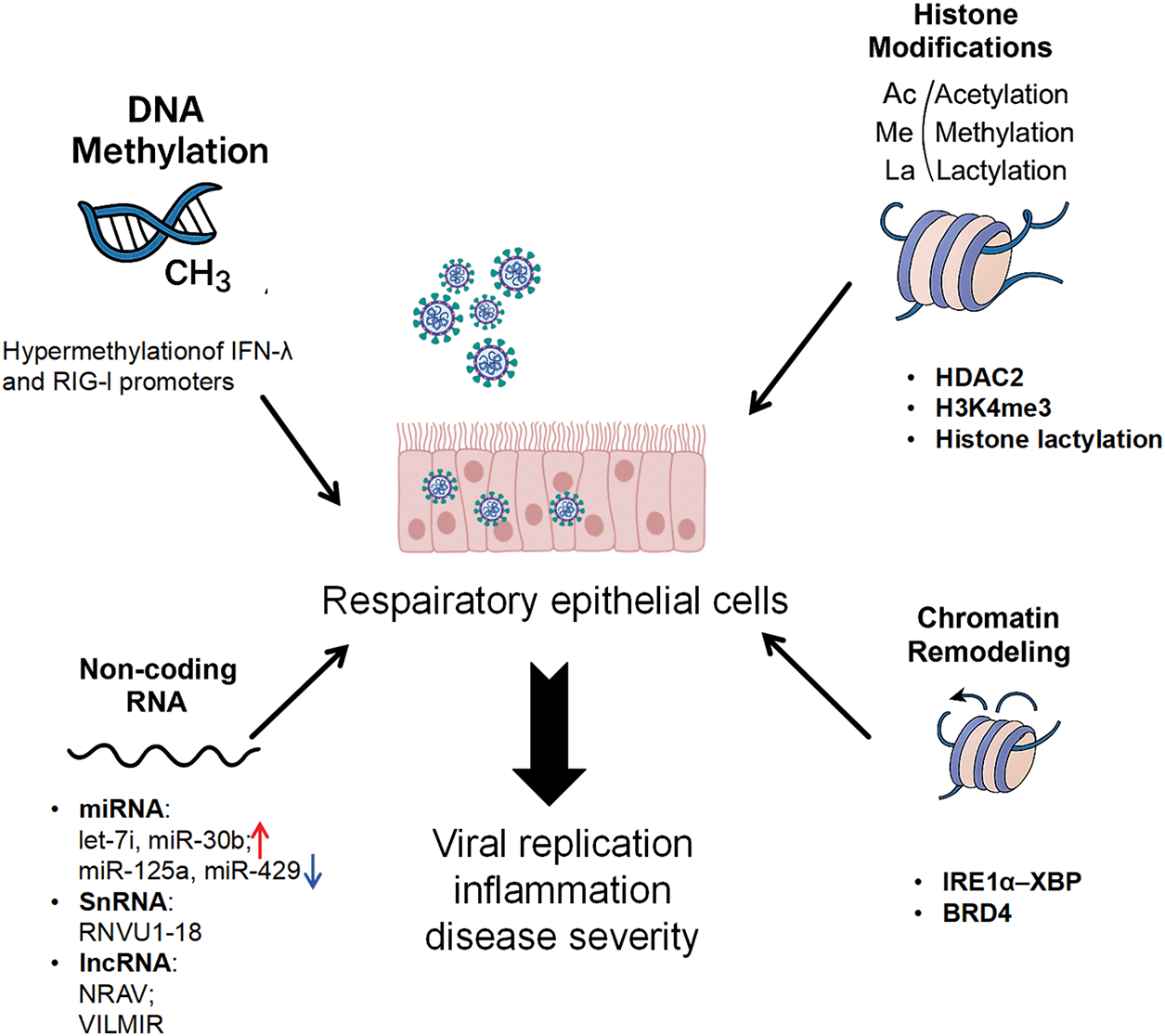
Figure 1. Epigenetic reprogramming of the airway epithelium during RSV infection. RSV reprograms its primary target, the respiratory epithelium, via four interconnected epigenetic axes that collectively govern viral replication, inflammation, and long-term airway pathology.
DNA Methylation: RSV can alter innate immune responses and airway pathology by modulating DNA methylation in respiratory epithelial cells. Similar to rhinovirus, which induces widespread CpG methylation changes in human nasal epithelial cells and activates DNA methyltransferases,56 RSV may also silence critical antiviral genes. Clinical epigenomic analyses of RSV-infected infants have identified differential host DNA methylation signatures associated with disease severity, consistent with epigenetic modulation of antiviral response pathways rather than random methylation changes.57 A longitudinal clinical study of children under 2 with RSV infection revealed that those who developed asthma or recurrent wheezing later exhibited significant differences in DNA methylation patterns compared to those who fully recovered, suggesting a link between methylation changes and long-term sequelae.58 Further, RSV immunoprophylaxis has been shown to alter the methylation profile of nasal epithelial cells in infants, indicating that both natural infection and preventive measures can epigenetically reprogram the airway cells.59 The clinical findings indicate that RSV and host responses dynamically and reversibly alter DNA methylation in airway epithelial cells, potentially influencing long-term antiviral immunity and respiratory health. While these methylation changes are consistently associated with childhood asthma and wheezing, they may serve as biomarkers for children at risk of these conditions.
Histone Modifications: RSV induces distinct histone modification patterns in respiratory epithelial cells, each differentially regulating antiviral and inflammatory gene expression. In human bronchial epithelial cells (BEAS-2B), RSV increases histone deacetylase 2 (HDAC2) expression. This reduces histone H3 acetylation (e.g., H3K9ac, H3K14ac) at promoters of antiviral genes such as IFN-β and pro-inflammatory genes such as IL-6 and IL-8. The resulting hypoacetylation suppresses transcription and weakens the innate immune response.60 Treatment with HDAC inhibitors, such as trihydroxypyridine or vorinostat, restores H3 acetylation, reactivates antiviral gene expression, and thus suppresses viral replication and inflammation, underscoring the protective role of histone acetylation during RSV infection. Conversely, in respiratory epithelial cells, RSV infection has been reported to induce enrichment of active chromatin marks (such as H3K4me3 and H3K9ac) at antiviral loci such as RIG-I, and studies in influenza A virus (IAV)–infected epithelial cell models further demonstrate that increased H3K4me3 at the promoters of IFN-β and IFN-stimulated genes (ISGs), including ISG15, augments RIG-I/mitochondrial antiviral-signaling protein–dependent type I IFN responses.61–63 The viral NS1 and NS2 proteins can counteract this by inhibiting host methyltransferases. This dual mechanism, suppressing immune gene expression through reduced acetylation while activating antiviral pathways through enhanced methylation, highlights the complex and dynamic regulation of histone marks during RSV infection. Understanding how these modifications are controlled across space and time is crucial for comprehending epithelial immune defense and for developing targeted epigenetic therapies for RSV.
Histone Lactylation: A newly discovered epigenetic modification, histone lactylation, is prevalent in various cell types and plays a crucial role in regulating gene transcription, metabolic processes, and immune responses.64 It influences inflammatory and immune responses by modifying histone–DNA interactions and transcription factor activity.65,66 In viral infections, lactylation can play a significant role in host–pathogen interactions; for instance, porcine reproductive and respiratory syndrome virus can increase histone lactylation to suppress IFN-β production.67,68 RSV infection induces metabolic reprogramming in respiratory epithelial cells in vitro, characterized by enhanced glucose uptake, lactate production, and upregulation of glycolytic enzymes like hexokinase 2.69 The accumulated lactate serves as a substrate for histone lactylation,70 positioning this modification as a potential mechanism for RSV to reprogram the epithelial epigenome, influencing both host antiviral immunity and inflammation. Investigating the role of histone lactylation represents a promising frontier in understanding RSV pathogenesis.
ncRNAs: RSV can also significantly alter epithelial cell function by regulating various ncRNAs.71 Clinical analyses of nasal swab specimens from RSV-infected infants identified distinct miRNA profiles, with miR-34b/c, miR-125b, miR-29c, miR-429, and miR-27b downregulated, and miR-155, miR-31, and let-7d upregulated.72 Low levels of miR-125a and miR-429 were associated with milder disease, suggesting a potential regulatory role of these miRNAs in influencing infection outcomes and their potential utility as biomarkers for disease progression. In bronchial epithelial models, RSV upregulates let-7i and miR-30b to suppress IFN signaling, while NS1 and NS2 proteins inhibit this upregulation to maintain immune balance.71 Beyond miRNAs, certain lncRNAs are involved in RSV pathogenesis. The pro-viral lncRNA, NRAV, promotes RSV replication by sequestering miR-509-3p, thereby enhancing Ras-related protein Rab-5C–mediated vesicular transport in infected cells in vitro.73 Conversely, the IFN-induced lncRNA, VILMIR, is upregulated upon infection and enhances antiviral gene transcription.74 The pseudogene-derived RNVU1-18, originating from U1 nucleolar RNA, is also upregulated and promotes RIG-I activation by interacting with tripartite motif-containing 25 (TRIM25) to facilitate K63-linked ubiquitination in vitro.75 This illustrates that ncRNAs form a precise, context-dependent regulatory network during RSV infection, with diverse and occasionally opposing functions governed by their expression timing and interactions with viral and host factors. A comprehensive understanding of the ncRNA landscape may unveil new biomarkers and therapeutic targets.
Chromatin Remodeling: RSV infection induces chromatin restructuring in respiratory epithelial cells, impacting antiviral responses and airway remodeling. Qiao et al. demonstrated, in human small airway epithelial cells in vitro and murine respirovirus lung infection models in vivo, that the activation of the unfolded protein response sensor inositol-dependent endonuclease 1α (IRE1α) and subsequent splicing of X-box binding protein 1s (XBP1s) increase chromatin accessibility at loci encoding hepatocyte-binding protein and epithelial–mesenchymal transition (EMT)-associated genes. This IRE1α–XBP1-driven remodeling boosts expression of antiviral and inflammatory genes and upregulates EMT programs, contributing to long-term pathology like asthma.76 Another in vitro study showed that RSV induces chromatin remodeling at the promoters of growth factors, such as transforming growth factor-beta (TGF-β), and extracellular matrix genes, including fibronectin-1 and matrix metalloproteinase 9, promoting epithelial proliferation and aberrant matrix deposition linked to airway narrowing and reduced lung function.77 Bromodomain-containing protein 4 (BRD4), a member of the bromodomain and extra-terminal family, also plays a critical role in these processes in human airway epithelial and basal cell models in vitro. By recognizing acetylated histone tails, BRD4 recruits transcription factors like positive transcription elongation factor b, which contains cyclin-dependent kinase 9, IRF1, and nuclear factor-kappa-light-chain-enhancer of activated B cells to the promoters of pro-inflammatory, antiviral, and remodeling-associated genes. This facilitates RNA polymerase II phosphorylation and efficient transcription elongation.78,79 These studies underscore that RSV-induced chromatin remodeling, mediated by pathways like IRE1α–XBP1s and BRD4, not only activates robust innate immunity but also predisposes airway epithelia to long-term structural changes, potentially underpinning the link to asthma.
Epigenetic Reprogramming of T Cell Differentiation and Immune Memory
T cells are central to adaptive immunity against RSV. CD8+ cytotoxic T lymphocytes directly eliminate infected cells and produce IFN-γ to aid viral clearance.80 Persistent tissue-resident memory CD8+ T cells in the lungs are crucial for protection against reinfection.81 However, excessive CD8+ T cell activation can cause immunopathology, as seen in mouse models where overproduction of tumor necrosis factor-α and IFN-γ exacerbates lung injury.82 Establishing a balanced CD8+ T cell response is crucial for effective defense against RSV.
CD4+ helper T cells orchestrate immune responses through cytokine secretion and are equally essential for RSV virus eradication. Th1 cells produce IFN-γ, which activates macrophages and enhances CD8+ T cell function,83 and helps B cells generate high-affinity antibodies.84 Dysregulated Th2 responses, however, drive immunopathology by recruiting eosinophils and promoting airway hyperresponsiveness.85 Th17 cells exacerbate damage through IL-17, which induces mucus overproduction and neutrophil infiltration.86 Regulatory T cells (Tregs) expand early in RSV infection and help limit tissue damage by secreting anti-inflammatory cytokines like TGF-β and IL-10.87
Epigenetic Mechanisms in T Cell Polarization: Epigenetic modifications critically determine T cell fate during RSV infection. RSV increases the expression of Nodal, a TGF-β superfamily ligand, by demethylating CpG sites in its promoter in bronchial epithelial cells in vitro. Elevated Nodal then drives naive CD4+ T cells toward Th2 and Th17 lineages in co-culture systems, worsening airway inflammation and remodeling.88 Histone modifications further shape T cell responses. RSV activates the Notch pathway through Delta-like ligand 4 on dendritic cells (DCs) in murine models of RSV infection and in Treg differentiation cultures, upregulating the histone methyltransferase MYND domain-containing protein 3 (SMYD3). SMYD3 catalyzes H3K4me3 at the Forkhead box protein 3 promoter and its conserved noncoding sequence 1 region, promoting Treg differentiation and enhancing their suppressive function.89 Conversely, RSV infection can also reduce levels of the repressive mark H3K27me3 in DCs through the demethylase lysine-specific demethylase 6 in bone marrow–derived DC (BM-DC) and pulmonary DC from RSV-infected mice, thereby alleviating the repression of Th2-promoting genes and facilitating Th2 polarization.90 Thus, DNA methylation and histone modifications collectively establish an RSV-regulated T cell landscape favoring both inflammatory (Th2/Th17) and Treg phenotypes, ultimately influencing pathology and disease outcomes (Fig. 2).
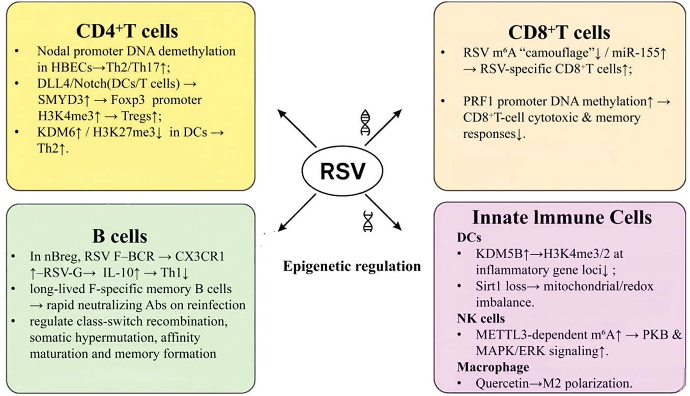
Figure 2. Epigenetic regulation of adaptive and innate immune responses to RSV infection. RSV infection induces a network of cell type–specific epigenetic alterations that collectively skew the immune response toward Th2 inflammation, impair cytotoxic memory, and elevate long-term asthma risk.
Fine-Tuning and Long-Term Effects: Epigenetic mechanisms also fine-tune specific T cell responses. N6-methyladenosine (m6A) modification of the RSV genome conceals viral pathogen-associated molecular patterns in vitro, helping the virus evade RIG-I detection. In mouse models, removal of this m6A “camouflage” enhances neutralizing antibody titers and RSV-specific T cell responses.91 ncRNAs, including miR-155, miR-146a, and the miR-17-92 cluster, modulate T cell activation, proliferation, differentiation, and cytokine production. For instance, miR-155 promotes viral clearance by enhancing Th1 polarization and CD8+ T cell effector function.92
RSV infection is associated with lasting epigenetic changes in T cells linked to allergic sensitization and immune memory. In a murine RSV infection model in vivo, RSV triggers demethylation of the Notch homolog 1, translocation-associated (Notch-1) promoter in CD4+ T cells, thereby enhancing the transcription of Th2/Th17 cytokines (such as IL-4, IL-13, and IL-17).93 This phenomenon may contribute to chronic airway inflammation and asthma susceptibility. Concurrently, RSV increases DNA methylation in the regulatory region of the perforin-1 gene in CD8+ T cells, which reduces perforin-1 expression. This epigenetic silencing impairs cytolytic activity and hinders the development of long-lived memory CD8+ T cells, thereby compromising durable antiviral protection.94 These coordinated epigenetic changes promote pathogenic CD4+ T cell responses while suppressing cytotoxic CD8+ T cell memory. Together, they create an immune environment skewed toward Th2 activity and reduced antiviral capacity. This imbalance favors chronic airway inflammation, airway remodeling, and ultimately increases the risk of asthma development.
Epigenetic Basis of B Cell Differentiation and Humoral Immunity
RSV infection significantly influences B cell differentiation and humoral immunity.95 Following infection, innate immune cells like neutrophils and macrophages initiate early antiviral responses,96 while antigen stimulation triggers naive B cells to differentiate into antibody-secreting plasma cells. RSV-specific antibodies neutralize free virus and persist in mucosal tissues and circulation, reducing reinfection risk.97 Specific B cell subsets exhibit distinct behaviors during RSV infection. For instance, binding of the RSV F protein to the B-cell receptor of natural regulatory B cells (nBreg) activates them, upregulating the chemokine receptor CX3CR1. Interaction between CX3CR1 and the RSV G protein facilitates nBreg infection, inducing IL-10 production that suppresses Th1 responses and contributes to the pathogenesis of severe bronchiolitis.98 RSV also elicits a robust memory B cell response in mucosa-associated lymphoid tissue. Shehata et al. demonstrated that RSV exposure generates long-lived memory B cells targeting the F protein, which, upon reinfection, produce potent neutralizing antibodies, providing valuable insights into persistent immunity and vaccine development.99
Potential Epigenetic Influences: There is no direct evidence linking epigenetic changes to B cell function in RSV infection. Our understanding is inferred from studies on other pathogens or general immunology. In non-RSV models, primarily mouse immunization systems in vivo with ex vivo B cell epigenomic profiling, antigen stimulation leaves an “experiential” epigenetic imprint on memory B cells in an IRF4-dependent manner. This epigenetic imprint progressively increases chromatin accessibility at loci that regulate plasma cell differentiation. Upon subsequent antigen exposure, this chromatin state influences B cell fate. Elevated B lymphocyte–induced maturation protein-1 expression promotes commitment to the plasma cell lineage. Conversely, increased activity of BTB and CNC homology 2 favors B cell re-entry into the germinal center rather than terminal differentiation.100 Insights from other viruses, such as IAV, show that memory B cells can exhibit sustained chromatin accessibility and transcriptional reprogramming, conferring “epigenetically primed” effector traits correlated with long-lived antibody responses.101 In a murine lymphocytic choriomeningitis virus infection model, early type I IFN signaling shapes the chromatin properties of memory B cells, and blocking the IFN receptor alters their subset composition, illustrating a mechanistic link between the infectious environment, cytokine signals, B cell epigenetics, and fate/functional preference.102 These findings suggest that analogous pathways possibly operate during RSV infection, though this requires experimental validation.
Nonviral studies confirm that epigenetic modifications regulate B cell processes, including class-switch recombination, somatic hypermutation, affinity maturation, and memory formation.103 Given the link between B cell dysfunction and severe RSV disease,104,105 we propose that RSV may alter B cell immunity through epigenetic mechanisms. Evidence from other viral and nonviral systems reveals that infection context and cytokine signaling can induce lasting chromatin and transcriptional changes in memory B cells. By extension, RSV infection may imprint similar epigenetic programs that influence B cell function and antibody durability. Future studies should apply longitudinal multi-omics approaches to B cells from RSV-infected humans or animal models. These approaches include single-cell ATAC sequencing, DNA methylation profiling, and histone mark analysis. When combined with functional assays, such as antibody repertoire sequencing, cytokine secretion profiling, and fate mapping after antigen reexposure, these data will enable mechanistic evaluation of whether epigenetic remodeling contributes to impaired humoral immunity and clinical outcomes in RSV infection.
Epigenetic Activation and Regulation of Innate Immune Cells
Innate immune cells, including DCs, natural killer (NK) cells, and macrophages, recognize RSV through pattern recognition receptors, triggering cytokine and IFN production.23,106 RSV infection also induces epigenetic changes in these cells, modulating chromatin accessibility and transcription factor recruitment to shape the course and outcome of the innate immune response.107,108
As bridges between innate and adaptive immunity, DCs undergo RSV-induced epigenetic changes. In RSV-infected BM-DCs, the expression of the histone demethylase lysine–specific demethylase 5B (KDM5B) increases. KDM5B removes activation-associated histone marks H3K4me3 and H3K4me2 from inflammatory gene loci. This decreases the transcription of inflammatory genes and, in DC-specific Kdm5b-deficient mice in vivo, prepares DCs to promote a Th 2-biased immune response.109 Conversely, loss of the NAD+-dependent deacetylase sirtuin-1 (Sirt1) in RSV-infected BM-DCs disrupts mitochondrial membrane potential and redox balance. This metabolic dysregulation is associated, in DC-specific Sirt1-deficient mice in vivo, with elevated proinflammatory cytokines (IL-1β, IL-6, IL-23) and reduced antiviral mediators (IL-12, IFN-β).110
NK cells are crucial effector cells that can rapidly eliminate virus-infected cells.111 Epigenetic mechanisms underpin their development, effector functions, and memory properties. m6A RNA methylation, catalyzed by methyltransferase-like 3 (METTL3), is essential for NK cell proliferation, survival, and cytotoxicity both in vivo and in vitro. During IL-15–driven maturation, METTL3-dependent m6A modification stabilizes Src-like domain 2–containing inositol 5-phosphatase-2 mRNA, activating PKB and mitogen-activated protein kinase/extracellular signal-regulated kinase signaling pathways to maintain NK cell homeostasis and function.112
Macrophages execute pathogen clearance through coordinated interactions with CD8+ T cells while preserving pulmonary immunological homeostasis by modulating lung inflammation. Evidence indicates that epigenetic mechanisms influence macrophage polarization during RSV infection. Quercetin, a compound known to modulate epigenetic pathways, including DNA methylation, histone modifications, and miRNA expression, promotes a shift of alveolar macrophages from a classically activated (M1) proinflammatory phenotype to an alternatively activated (M2) anti-inflammatory phenotype both in vitro and in vivo. In RSV-infected mice, this shift markedly reduces lung inflammation.113 This indicates that epigenetic regulation of macrophage polarization may influence RSV disease severity.
In summary, RSV employs diverse epigenetic strategies to alter the function of innate immune cells, fine-tuning cytokine production, cellular polarization, and maturation processes to establish an immunological equilibrium that often favors viral persistence or immunopathology (Fig. 2).
Translational Prospects and Challenges
Epigenetic Biomarkers for RSV Severity and Prognosis
The current prevention and treatment of RSV face numerous challenges. Preventive strategies primarily rely on monoclonal antibodies, specifically palivizumab and nirsevimab, while various vaccination options,114 including mRNA-based formulations, are in clinical trials. Infants and young children are particularly susceptible to severe RSV illness due to their underdeveloped immune systems, narrow airways, and excessive secretions, which may sometimes lead to hospitalization and fatal outcomes. Further, accurate classification of disease severity is essential for optimizing treatment and prognosis. RSV-induced epigenetic alterations in inflammatory and antiviral pathways are linked to clinical severity and long-term outcomes, making them promising biomarker candidates.58,115 Cohort studies have identified sustained hypomethylation at CpG sites within the zinc finger and BTB domain–containing 38 and TRIM6–TRIM34 loci in infants with severe RSV bronchiolitis, suggesting that these patterns may predict disease progression.57 Elevated miR-155 levels in blood and sputum from severe RSV cases also highlight its potential as an early warning sign for high-risk individuals.116 Integrating such epigenetic markers into diagnostic and prognostic workflows could enable earlier interventions and improve clinical outcomes.
Epigenetic Therapeutics: HDAC Inhibitors, CRISPR Tools, and Beyond
Epigenetic pathways, which can govern all stages of RSV infection and replication, represent attractive therapeutic targets. Strategies that modulate host chromatin regulators can inhibit viral replication, potentiate antiviral defenses, and enhance viral clearance, potentially overcoming challenges posed by the high antigenic variability of viral proteins, such as the RSV F protein. HDAC inhibitors can modulate antiviral responses by increasing histone acetylation at select antiviral gene promoters, although these effects depend on enzyme, cell type, and context.117,118 HDAC inhibition has been shown to reduce RSV replication in vitro and alleviate airway inflammation in mouse models, underscoring its therapeutic potential.60 Clustered Regularly Interspaced Short Palindromic Repeats (CRISPR)-associated protein 9 (CRISPR/Cas9) technology, a versatile gene-editing platform,119,120 has been applied to engineer B cells to produce RSV-neutralizing antibodies and to directly cleave persistent viral genomes, such as hepatitis B virus and human immunodeficiency virus, within host cells, thereby inhibiting viral replication.121,122 Looking ahead, CRISPR/Cas9 could be harnessed to establish a sustained antiviral state by epigenetically modifying host cells, particularly airway epithelial or immune cells, through targeted regulation of key epigenetic enzymes such as HDACs. Despite its potential, CRISPR/Cas9 needs further refinement to reduce off-target effects and improve delivery efficiency. While cancer, frequently propelled by epigenetic dysregulation, has been the primary focus of CRISPR/Cas9 therapy, substantial technical obstacles persist.123
Challenges, Safety, and Ethical Considerations
The advancement of epigenetic therapies for RSV encounters multiple scientific and translational hurdles. A central methodological challenge arises from the inherent cell-type- and developmental-stage-specificity of epigenetic modifications, which exhibit complex and evolving spatiotemporal dynamics during infection.124 Comprehensive mapping of these dynamics requires sophisticated high-throughput methodologies, imposing significant technical and financial burdens. This challenge is compounded by the field’s traditional reliance on in vitro systems and murine models, which frequently fail to capture key nuances of human-specific immune responses. To address this gap, recent research has increasingly shifted toward more physiologically relevant human models, such as airway organoids and precision-cut lung slices, which facilitate more accurate translational predictions.125,126
Equally critical are the safety concerns associated with epigenetic modulators, with off-target effects representing a primary obstacle to clinical development. Many candidate agents, including broad-spectrum HDAC inhibitors, exhibit pleiotropic activity that can disrupt transcriptional homeostasis in healthy, uninfected tissues.127,128 This risk is markedly amplified in pediatric populations. Infants and young children undergo a phase of exceptionally rapid physiological development, rendering their organ systems uniquely vulnerable to developmental toxicities. Preclinical evidence substantiates this concern, indicating that systemic HDAC inhibition can impair osteoblast maturation and chondrocyte function, thereby posing a tangible risk of skeletal growth impairment—a particularly serious potential adverse outcome in this age group.129,130 Further complicating the safety profile is the well-established role of HDACs in regulating synaptic plasticity, which raises the possibility of unintended neurodevelopmental consequences—an unresolved issue that necessitates vigilant long-term monitoring. Consequently, establishing a viable therapeutic window for pediatric applications will likely require the development of next-generation inhibitors with enhanced selectivity or the implementation of sophisticated targeted delivery platforms.
The prospective clinical translation of genome-editing technologies such as CRISPR/Cas9 introduces additional layers of ethical and regulatory complexity. A paramount concern, both scientifically and ethically, is the irreversible nature of heritable genetic edits. In the context of actively proliferating pediatric tissues, even rare off-target mutations may undergo clonal expansion, potentially conferring latent oncogenic risks that might not manifest until adulthood.131 From a regulatory and ethical standpoint, designing first-in-human trials for infant cohorts presents the dual challenge of securing valid informed consent and ensuring a justifiable risk-benefit ratio. Prevailing ethical frameworks typically stipulate that any research risk exceeding a minimal threshold must be counterbalanced by a clear prospect of direct therapeutic benefit for the participant.132 Therefore, future clinical protocols must incorporate stringent and prolonged follow-up strategies, designed not only to assess treatment efficacy but also to monitor long-term genomic stability and the potential emergence of delayed adverse events throughout the lifespan.
Future Avenues: Multi-Omics Integration and Organoid Models
While contemporary epigenetic research continues to rely predominantly on bulk cell analyses or immortalized lines, a clear shift is underway toward high-resolution models with direct patient relevance. Integrating multi-omics—spanning epigenomics, transcriptomics, and metabolomics—with human airway organoids now defines the cutting edge of RSV investigation.133–137 In contrast to conventional two-dimensional cultures, human nasal organoids (HNOs) generated from patient samples faithfully recreate the multicellular architecture, functional ciliary activity, and mucus secretion characteristic of the native airway epithelium.138,139 This makes them a powerful experimental system for probing age-specific host–virus dynamics.140
Emerging primary research underscores the transformative impact of such models. For example, Rajan et al. applied single-cell RNA sequencing—a technique well suited for resolving cellular heterogeneity—to profile RSV-infected HNOs derived from pediatric and adult donors. Their work uncovered pronounced age-dependent differences in cellular responses, including a distinct “proliferative diversity” unique to infant tissues. Moreover, the study revealed an extended cellular tropism of RSV in infants, with the virus targeting a wider array of epithelial cell types—a finding that aligns with the increased clinical severity observed in this age group.141 Complementing this, Aloisio et al. utilized infant-derived HNOs to demonstrate that pediatric epithelium displays significantly higher susceptibility to RSV infection, along with more vigorous inflammatory responses and greater cytotoxicity, compared to adult-derived counterparts.142
Together, these observations highlight the unique value of organoid-based systems combined with single-cell methodologies. Moving forward, epigenetic studies should capitalize on these models to map features such as chromatin accessibility and histone modifications at single-cell resolution. Achieving this level of detail will be crucial for determining whether a persistent “epigenetic memory” of RSV infection resides within specific progenitor cell populations. This mechanism could ultimately explain long-term clinical sequelae, including recurrent wheezing.
Conclusion
This review emphasizes the key role of host epigenetic reprogramming in shaping immune responses and affecting viral replication during RSV infection. RSV manipulates multiple layers of epigenetic regulation, such as DNA methylation, histone modifications, ncRNAs, and chromatin remodeling, across respiratory epithelial cells, T cells, B cells, and innate immune populations. These changes interfere with normal gene expression and influence clinical outcomes, from acute disease severity to long-term respiratory issues like childhood asthma. Although emerging epigenetic-based therapies show promise, further research is needed. Combining multi-omics approaches with advanced human-relevant systems, such as patient-derived airway organoids, will be crucial to fully understand the epigenetic networks driving RSV pathogenesis and to accelerate the development of more targeted therapeutics and vaccines.
Acknowledgment
We are grateful to our collaborators and colleagues who provided valuable insights and feedback throughout the study. Finally, we extend our thanks to the editors and reviewers for their constructive comments that helped improve the quality of our manuscript.
Funding Statement
This study was funded by the National Natural Science Foundation of China (Nos. 82200011 and 82401989), the China Postdoctoral Science Foundation (No. 2025M772162), and the Science and Technology Research Program Project of Chongqing Municipal Education Commission (Nos. KJQN202402815, KJQN202302818).
Author Contributions
G. Y., M. W., and H. F. contributed to the conception and design of the study and to drafting the manuscript. Y. L., S. C., J. L., and Z. L. were responsible for the revision and critical review of the article. All authors have agreed on the journal to which the article has been submitted and accept responsibility for all aspects of the work.
Data Availability Statement
No additional data was necessary for the manuscript.
Ethics Approval Statement
Not applicable.
Conflict of Interest Disclosure
The authors declare that they have no competing interests.
Supplemental Information
Supplemental information for this article can be found online at https://sup.jclinque.com/api/articles/100/download-suppl.
References
[1] Swedan S, Andrews J, Majumdar T, Musiyenko A, Barik S. Multiple functional domains and complexes of the two nonstructural proteins of human respiratory syncytial virus contribute to interferon suppression and cellular location. J Virol. 2011;85(19):10090–10100. doi:10.1128/JVI.00413-11.
[2] McLellan JS, Ray WC, Peeples ME. Structure and function of respiratory syncytial virus surface glycoproteins. Curr Top Microbiol Immunol. 2013;372:83–104. doi:10.1007/978-3-642-38919-1_4.
[3] Schwarze J, Schauer U. Enhanced virulence, airway inflammation and impaired lung function induced by respiratory syncytial virus deficient in secreted G protein. Thorax. 2004;59(6):517–521. doi:10.1136/thx.2003.017343.
[4] Melero JA, Mas V, McLellan JS. Structural, antigenic and immunogenic features of respiratory syncytial virus glycoproteins relevant for vaccine development. Vaccine. 2017;35(3):461–468. doi:10.1016/j.vaccine.2016.09.045.
[5] Huong TN, Ravi Iyer L, Lui J, Wang DY, Tan BH, Sugrue RJ. The respiratory syncytial virus SH protein is incorporated into infectious virus particles that form on virus-infected cells. Virology. 2023;580:28–40. doi:10.1016/j.virol.2023.01.013.
[6] Sibert BS, Kim JY, Yang JE, et al. Assembly of respiratory syncytial virus matrix protein lattice and its coordination with fusion glycoprotein trimers. Nat Commun. 2024;15(1):5923. doi:10.1038/s41467-024-50162-x.
[7] Wang Y, Zhang C, Luo Y, et al. Cryo-EM structure of the nucleocapsid-like assembly of respiratory syncytial virus. Signal Transduct Target Ther. 2023;8(1):323. doi:10.1038/s41392-023-01602-5.
[8] Asenjo A, Mendieta J, Gómez-Puertas P, Villanueva N. Residues in human respiratory syncytial virus P protein that are essential for its activity on RNA viral synthesis. Virus Res. 2008;132(1-2):160–173. doi:10.1016/j.virusres.2007.11.013.
[9] Cadena-Cruz C, Villarreal Camacho JL, De Ávila-Arias M, et al. Respiratory syncytial virus entry mechanism in host cells: a general overview. Mol Microbiol. 2023;120(3):341–350. doi:10.1111/mmi.15133.
[10] Hayes RS, Oraby AK, Camargo C, Marchant DJ, Sagan SM. Mapping respiratory syncytial virus fusion protein interactions with the receptor IGF1R and the impact of alanine-scanning mutagenesis on viral infection. J Gen Virol. 2024;105(1):001951. doi:10.1099/jgv.0.001951.
[11] Gilman MSA, Liu C, Fung A, et al. Structure of the respiratory syncytial virus polymerase complex. Cell. 2019;179(1):193–204.e14. doi:10.1016/j.cell.2019.08.014.
[12] Kurt-Jones EA, Popova L, Kwinn L, et al. Pattern recognition receptors TLR4 and CD14 mediate response to respiratory syncytial virus. Nat Immunol. 2000;1(5):398–401. doi:10.1038/80833.
[13] Kolli D, Velayutham TS, Casola A. Host-viral interactions: role of pattern recognition receptors (PRRs) in human pneumovirus infections. Pathogens. 2013;2(2):232–263. doi:10.3390/pathogens2020232.
[14] Sharma P, Hoorn D, Aitha A, Breier D, Peer D. The immunostimulatory nature of mRNA lipid nanoparticles. Adv Drug Deliv Rev. 2024;205:115175. doi:10.1016/j.addr.2023.115175.
[15] Guo X, Liu T, Shi H, et al. Respiratory syncytial virus infection upregulates NLRC5 and major histocompatibility complex class I expression through RIG-I induction in airway epithelial cells. J Virol. 2015;89(15):7636–7645. doi:10.1128/JVI.00349-15.
[16] Chaudhary R, Meher A, Krishnamoorthy P, Kumar H. Interplay of host and viral factors in inflammatory pathway mediated cytokine storm during RNA virus infection. Curr Res Immunol. 2023;4:100062. doi:10.1016/j.crimmu.2023.100062.
[17] Chirkova T, Boyoglu-Barnum S, Gaston KA, et al. Respiratory syncytial virus G protein CX3C motif impairs human airway epithelial and immune cell responses. J Virol. 2013;87(24):13466–13479. doi:10.1128/JVI.01741-13.
[18] Anderson J, Do LAH, van Kasteren PB, Licciardi PV. The role of respiratory syncytial virus G protein in immune cell infection and pathogenesis. EBioMedicine. 2024;107:105318. doi:10.1016/j.ebiom.2024.105318.
[19] Obando-Pacheco P, Justicia-Grande AJ, Rivero-Calle I, et al. Respiratory syncytial virus seasonality: a global overview. J Infect Dis. 2018;217(9):1356–1364. doi:10.1093/infdis/jiy056.
[20] Agac A, Kolbe SM, Ludlow M, Osterhaus ADME, Meineke R, Rimmelzwaan GF. Host responses to respiratory syncytial virus infection. Viruses. 2023;15(10):1999. doi:10.3390/v15101999.
[21] Li Y, Wang X, Blau DM, et al. Global, regional, and national disease burden estimates of acute lower respiratory infections due to respiratory syncytial virus in children younger than 5 years in 2019: a systematic analysis. Lancet. 2022;399(10340):2047–2064. doi:10.1016/S0140-6736(22)00478-0.
[22] Pandya MC, Callahan SM, Savchenko KG, Stobart CC. A contemporary view of respiratory syncytial virus (RSV) biology and strain-specific differences. Pathogens. 2019;8(2):67. doi:10.3390/pathogens8020067.
[23] Marr N, Turvey SE, Grandvaux N. Pathogen recognition receptor crosstalk in respiratory syncytial virus sensing: a host and cell type perspective. Trends Microbiol. 2013;21(11):568–574. doi:10.1016/j.tim.2013.08.006.
[24] Sutto-Ortiz P, Eléouët J-F, Ferron F, Decroly E. Biochemistry of the respiratory syncytial virus L protein embedding RNA polymerase and capping activities. Viruses. 2023;15(2):341. doi:10.3390/v15020341.
[25] Branche AR, Falsey AR. Respiratory syncytial virus infection in older adults: an under-recognized problem. Drugs & Aging. 2015;32(4):261–269. doi:10.1007/s40266-015-0258-9.
[26] Robinson JL, Papenburg J. An update on prevention of paediatric respiratory syncytial virus hospitalizations in Canada. J Assoc Med Microbiol Infect Dis Can. 2025;10(1):2–5. doi:10.3138/jammi-2025-0203.
[27] Fitzpatrick T, Buchan SA, Mahant S, et al. Pediatric respiratory syncytial virus hospitalizations, 2017–2023. JAMA Netw Open. 2024;7(6):e2416077. doi:10.1001/jamanetworkopen.2024.16077.
[28] Groves HE, Piché-Renaud PP, Peci A, et al. The impact of the COVID-19 pandemic on influenza, respiratory syncytial virus, and other seasonal respiratory virus circulation in Canada: a population-based study. Lancet Reg Health Am. 2021;1:100015. doi:10.1016/j.lana.2021.100015.
[29] Otomaru H, Sornillo JBT, Kamigaki T, et al. Risk of transmission and viral shedding from the time of infection for respiratory syncytial virus in households. Am J Epidemiol. 2021;190(12):2536–2543. doi:10.1093/aje/kwab181.
[30] Löwensteyn YN, Willemsen JE, Mazur NI, et al. Nosocomial RSV-related in-hospital mortality in children <5 years: a global case series. Pediatr Infect Dis J. 2023;42(1):1–7. doi:10.1097/INF.0000000000003747.
[31] Aujard Y, Fauroux B. Risk factors for severe respiratory syncytial virus infection in infants. Respir Med. 2002;96 Suppl B:S9–S14. doi:10.1053/rmed.2002.1295.
[32] Tahamtan A, Askari FS, Bont L, Salimi V. Disease severity in respiratory syncytial virus infection: role of host genetic variation. Rev Med Virol. 2019;29(2):e2026. doi:10.1002/rmv.2026.
[33] Khawaja F, Chemaly RF. Respiratory syncytial virus in hematopoietic cell transplant recipients and patients with hematologic malignancies. Haematologica. 2019;104(7):1322–1331. doi:10.3324/haematol.2018.215152.
[34] Herrmann S, Graefe S, Christopeit M, et al. Respiratory syncytial virus infection in patients with haematological diseases: a retrospective multicentre study. Infection. 2025;53(4):1341–1350. doi:10.1007/s15010-024-02449-w.
[35] Wildenbeest JG, Lowe DM, Standing JF, Butler CC. Respiratory syncytial virus infections in adults: a narrative review. Lancet Respir Med. 2024;12(10):822–836. doi:10.1016/S2213-2600(24)00255-8.
[36] Wang X, Li Y, Shi T, et al. Global disease burden of and risk factors for acute lower respiratory infections caused by respiratory syncytial virus in preterm infants and young children in 2019: a systematic review and meta-analysis of aggregated and individual participant data. Lancet. 2024;403(10433):1241–1253. doi:10.1016/S0140-6736(24)00138-7.
[37] O’Brien KL, Baggett HC, Brooks WA, et al. Causes of severe pneumonia requiring hospital admission in children without HIV infection from Africa and Asia: the PERCH multi-country case-control study. Lancet. 2019;394(10200):757–779. doi:10.1016/S0140-6736(19)30721-4.
[38] Wildenbeest JG, Billard MN, Zuurbier RP, et al. The burden of respiratory syncytial virus in healthy term-born infants in Europe: a prospective birth cohort study. Lancet Respir Med. 2023;11(4):341–353. doi:10.1016/S2213-2600(22)00414-3.
[39] Perez A, Lively JY, Curns A, et al. Respiratory virus surveillance among children with acute respiratory illnesses - new vaccine surveillance network, United States, 2016–2021. MMWR Morb Mortal Wkly Rep. 2022;71(40):1253–1259. doi:10.15585/mmwr.mm7140a1.
[40] Blau DM, Baillie VL, Els T, et al. Deaths attributed to respiratory syncytial virus in young children in high-mortality rate settings: report from child health and mortality prevention surveillance (CHAMPS). Clin Infect Dis. 2021;73(Suppl_3):S218–S228. doi:10.1093/cid/ciab509.
[41] Scheltema NM, Gentile A, Lucion F, et al. Global respiratory syncytial virus-associated mortality in young children (RSV GOLD): a retrospective case series. Lancet Glob Health. 2017;5(10):e984–e991. doi:10.1016/S2214-109X(17)30344-3.
[42] Biggs HM, Simões EAF, Abu Khader I, et al. Respiratory syncytial virus infection among hospitalized infants in four middle-income countries. J Pediatric Infect Dis Soc. 2023;12(7):394–405. doi:10.1093/jpids/piad042.
[43] Soni A, Kabra SK, Lodha R. Respiratory syncytial virus infection: an update. Indian J Pediatr. 2023;90(12):1245–1253. doi:10.1007/s12098-023-04613-w.
[44] Parums DV. Editorial: Surveillance of seasonal respiratory syncytial virus (RSV) infection in children and vulnerable adults drives vaccine development and new immunization programs. Med Sci Monit. 2025;31:e949558. doi:10.12659/MSM.949558.
[45] Zar HJ, Simões EAF, Madhi SA, et al. Clesrovimab for prevention of RSV disease in healthy infants. N Engl J Med. 2025;393(13):1292–1303. doi:10.1056/NEJMoa2502984.
[46] Wang L, Tang J. SWI/SNF complexes and cancers. Gene. 2023;870:147420. doi:10.1016/j.gene.2023.147420.
[47] Chen HS, Wang F, Chen JG. Epigenetic mechanisms in depression: implications for pathogenesis and treatment. Curr Opin Neurobiol. 2024;85:102854. doi:10.1016/j.conb.2024.102854.
[48] Izadi M, Sadri N, Abdi A, Serajian S, Jalalei D, Tahmasebi S. Epigenetic biomarkers in aging and longevity: current and future application. Life Sci. 2024;351:122842. doi:10.1016/j.lfs.2024.122842.
[49] Moore LD, Le T, Fan G. DNA methylation and its basic function. Neuropsychopharmacology. 2013;38(1):23–38. doi:10.1038/npp.2012.112.
[50] Zu H, Chen X. Epigenetics behind CD8(+) T cell activation and exhaustion. Genes Immun. 2024;25(6):525–540. doi:10.1038/s41435-024-00307-1.
[51] Rajanathadurai J, Perumal E, Sindya J. Advances in targeting cancer epigenetics using CRISPR-dCas9 technology: a comprehensive review and future prospects. Funct Integr Genomics. 2024;24(5):164. doi:10.1007/s10142-024-01455-3.
[52] Elkhalifa AME, Nabi SU, Shah OS, et al. Insight into oncogenic viral pathways as drivers of viral cancers: implication for effective therapy. Curr Oncol. 2023;30(2):1924–1944. doi:10.3390/curroncol30020150.
[53] Mattick JS, Makunin IV. Non-coding RNA. Hum Mol Genet. 2006;15 Spec No 1:R17–29. doi:10.1093/hmg/ddl046.
[54] Clapier CR, Cairns BR. The biology of chromatin remodeling complexes. Annu Rev Biochem. 2009;78(1):273–304. doi:10.1146/annurev.biochem.77.062706.153223.
[55] Villenave R, Shields MD, Power UF. Respiratory syncytial virus interaction with human airway epithelium. Trends Microbiol. 2013;21(5):238–244. doi:10.1016/j.tim.2013.02.004.
[56] Pech M, Weckmann M, König IR, et al. Rhinovirus infections change DNA methylation and mRNA expression in children with asthma. PLoS One. 2018;13(11):e0205275. doi:10.1371/journal.pone.0205275.
[57] Pischedda S, Gómez-Carballa A, Pardo-Seco J, et al. DNA methylation signatures of severe RSV infection in infants: evidence from non-invasive saliva samples. Epigenetics Chromatin. 2025;18(1):25. doi:10.1186/s13072-025-00587-5.
[58] Pischedda S, Rivero-Calle I, Gómez-Carballa A, et al. Role and diagnostic performance of host epigenome in respiratory morbidity after RSV infection: the EPIRESVi study. Front Immunol. 2022;13:875691. doi:10.3389/fimmu.2022.875691.
[59] Xu CJ, Scheltema NM, Qi C, et al. Infant RSV immunoprophylaxis changes nasal epithelial DNA methylation at 6 years of age. Pediatr Pulmonol. 2021;56(12):3822–3831. doi:10.1002/ppul.25643.
[60] Feng Q, Su Z, Song S, et al. Histone deacetylase inhibitors suppress RSV infection and alleviate virus-induced airway inflammation. Int J Mol Med. 2016;38(3):812–822. doi:10.3892/ijmm.2016.2691.
[61] Spalluto CM, Singhania A, Cellura D, et al. IFN-γ influences epithelial antiviral responses via histone methylation of the RIG-I promoter. Am J Respir Cell Mol Biol. 2017;57(4):428–438. doi:10.1165/rcmb.2016-0392OC.
[62] Zhao L, Xia M, Wang K, et al. A long non-coding RNA IVRPIE promotes host antiviral immune responses through regulating interferon β1 and ISG expression. Front Microbiol. 2020;11:260. doi:10.3389/fmicb.2020.00260.
[63] Ji X, Meng W, Liu Z, Mu X. Emerging roles of lncRNAs regulating RNA-mediated type-I interferon signaling pathway. Front Immunol. 2022;13:811122. doi:10.3389/fimmu.2022.811122.
[64] Meng F, He J, Zhang X, et al. Histone lactylation antagonizes senescence and skeletal muscle aging by modulating aging-related pathways. Adv Sci (Weinh). 2025;12(22):2412747. doi:10.1002/advs.202412747.
[65] Zhang Y, Zhang X. Virus-induced histone lactylation promotes virus infection in crustacean. Adv Sci (Weinh). 2024;11(30):e2401017. doi:10.1002/advs.202401017.
[66] He Y, Huang Y, Peng P, Yan Q, Ran L. Lactate and lactylation in gastrointestinal cancer: current progress and perspectives (Review). Oncol Rep. 2024;53(1):6. doi:10.3892/or.2024.8839.
[67] Pang Y, Zhou Y, Wang Y, et al. Lactate-lactylation-HSPA6 axis promotes PRRSV replication by impairing IFN-β production. Gallagher T, ed. J Virol. 2024;98(1):e01670-23. doi:10.1128/jvi.01670-23.
[68] Chen S, Qin T, Luo S, et al. Lactylation and viral infections: a novel link between metabolic reprogramming and immune regulation. Taylor H, ed. PLoS Pathog. 2025;21(7):e1013366. doi:10.1371/journal.ppat.1013366.
[69] Morris DR, Qu Y, Agrawal A, Garofalo RP, Casola A. HIF-1α modulates core metabolism and virus replication in primary airway epithelial cells infected with respiratory syncytial virus. Viruses. 2020;12(10):1088. doi:10.3390/v12101088.
[70] Chen AN, Luo Y, Yang YH, et al. Lactylation, a novel metabolic reprogramming code: current status and prospects. Front Immunol. 2021;12:688910. doi:10.3389/fimmu.2021.688910.
[71] Thornburg NJ, Hayward SL, Crowe JE Jr. Respiratory syncytial virus regulates human microRNAs by using mechanisms involving beta interferon and NF-κB. mBio. 2012;3(6):e00220-12. doi:10.1128/mBio.00220-12.
[72] Inchley CS, Sonerud T, Fjærli HO, Nakstad B. Nasal mucosal microRNA expression in children with respiratory syncytial virus infection. BMC Infect Dis. 2015;15(1):150. doi:10.1186/s12879-015-0878-z.
[73] Li J, Li M, Wang X, et al. Long noncoding RNA NRAV promotes respiratory syncytial virus replication by targeting the MicroRNA miR-509-3p/Rab5c axis to regulate vesicle transportation. J Virol. 2020;94(10):e00113-20. doi:10.1128/JVI.00113-20.
[74] John K, Huntress I, Smith E, et al. Human long noncoding RNA VILMIR is induced by major respiratory viral infections and modulates the host interferon response. Gallagher T, ed. J Virol. 2025;99(4):e00141-25. doi:10.1128/jvi.00141-25.
[75] Zhang F, Liu S, Qiao Z, et al. Housekeeping U1 snRNA facilitates antiviral innate immunity by promoting TRIM25-mediated RIG-I activation. Cell Rep. 2024;43(3):113945. doi:10.1016/j.celrep.2024.113945.
[76] Qiao D, Skibba M, Xu X, Garofalo RP, Zhao Y, Brasier AR. Paramyxovirus replication induces the hexosamine biosynthetic pathway and mesenchymal transition via the IRE1α-XBP1s arm of the unfolded protein response. Am J Physiol Lung Cell Mol Physiol. 2021;321(3):L576–L594. doi:10.1152/ajplung.00127.2021.
[77] Xu X, Qiao D, Mann M, Garofalo RP, Brasier AR. Respiratory syncytial virus infection induces chromatin remodeling to activate growth factor and extracellular matrix secretion pathways. Viruses. 2020;12(8):804. doi:10.3390/v12080804.
[78] Brasier AR. RSV reprograms the CDK9•BRD4 chromatin remodeling complex to couple innate inflammation to airway remodeling. Viruses. 2020;12(4):472. doi:10.3390/v12040472.
[79] Xu X, Qiao D, Brasier AR. Cooperative interaction of interferon regulatory factor-1 and bromodomain-containing protein 4 on RNA polymerase activation for intrinsic innate immunity. Front Immunol. 2024;15:1366235. doi:10.3389/fimmu.2024.1366235.
[80] De C, Pickles RJ, Yao W, et al. Human T cells efficiently control RSV infection. J Clin Investigat Insight. 2023;8(11):e168110. doi:10.1172/jci.insight.168110.
[81] Retamal-Díaz A, Covián C, Pacheco GA, et al. Contribution of resident memory CD8+ T cells to protective immunity against respiratory syncytial virus and their impact on vaccine design. Pathogens. 2019;8(3):147. doi:10.3390/pathogens8030147.
[82] Schmidt ME, Varga SM. The CD8 T cell response to respiratory virus infections. Front Immunol. 2018;9:678. doi:10.3389/fimmu.2018.00678.
[83] Kervevan J, Chakrabarti LA. Role of CD4+ T cells in the control of viral infections: recent advances and open questions. Int J Mol Sci. 2021;22(2):523. doi:10.3390/ijms22020523.
[84] Swain SL, McKinstry KK, Strutt TM. Expanding roles for CD4+ T cells in immunity to viruses. Nat Rev Immunol. 2012;12(2):136–148. doi:10.1038/nri3152.
[85] Knudson CJ, Hartwig SM, Meyerholz DK, et al. RSV vaccine-enhanced disease is orchestrated by the combined actions of distinct CD4 T cell subsets. Thomas PG, ed. PLoS Pathog. 2015;11(3):e1004757. doi:10.1371/journal.ppat.1004757.
[86] Cheon IS, Kim JY, Choi Y, et al. Sublingual immunization with an RSV G glycoprotein fragment primes IL-17-mediated immunopathology upon respiratory syncytial virus infection. Front Immunol. 2019;10:567. doi:10.3389/fimmu.2019.00567.
[87] Peng W, Wang L, Zhang H, Zhang Z, Chen X. Effects of recombinant IL-35-BCG on Treg/Th17 cell imbalance and inflammatory response in asthmatic newborn mice induced by RSV. Inflammation. 2021;44(6):2476–2485. doi:10.1007/s10753-021-01517-9.
[88] Wang L, Wu G, Qin X, et al. Expression of nodal on bronchial epithelial cells influenced by lung microbes through DNA methylation modulates the differentiation of T-helper cells. Cell Physiol Biochem. 2015;37(5):2012–2022. doi:10.1159/000438561.
[89] Ting HA, de Almeida Nagata D, Rasky AJ, et al. Notch ligand Delta-like 4 induces epigenetic regulation of Treg cell differentiation and function in viral infection. Mucosal Immunol. 2018;11(5):1524–1536. doi:10.1038/s41385-018-0052-1.
[90] Malinczak CA, Rasky AJ, Fonseca W, et al. Upregulation of H3K27 demethylase KDM6 during respiratory syncytial virus infection enhances proinflammatory responses and immunopathology. J Immunol. 2020;204(1):159–168. doi:10.4049/jimmunol.1900741.
[91] Xue M, Zhang Y, Wang H, et al. Viral RNA N6-methyladenosine modification modulates both innate and adaptive immune responses of human respiratory syncytial virus. PLoS Pathog. 2021;17(12):e1010142. doi:10.1371/journal.ppat.1010142.
[92] Głobińska A, Pawełczyk M, Kowalski ML. MicroRNAs and the immune response to respiratory virus infections. Expert Rev Clin Immunol. 2014;10(7):963–971. doi:10.1586/1744666X.2014.913482.
[93] Qin L, Qiu K, Hu C, Wang L, Wu G, Tan Y. Respiratory syncytial virus promoted the differentiation of Th17 cells in airway microenvironment through activation of Notch-1/Delta3. J Med Microbiol. 2019;68(4):649–656. doi:10.1099/jmm.0.000959.
[94] Elgizouli M, Logan C, Grychtol R, Rothenbacher D, Nieters A, Heinzmann A. Reduced PRF1 enhancer methylation in children with a history of severe RSV bronchiolitis in infancy: an association study. BMC Pediatr. 2017;17(1):65. doi:10.1186/s12887-017-0817-9.
[95] Domachowske JB, Rosenberg HF. Respiratory syncytial virus infection: immune response, immunopathogenesis, and treatment. Clin Microbiol Rev. 1999;12(2):298–309. doi:10.1128/CMR.12.2.298.
[96] Russell CD, Unger SA, Walton M, Schwarze J. The human immune response to respiratory syncytial virus infection. Clin Microbiol Rev. 2017;30(2):481–502. doi:10.1128/CMR.00090-16.
[97] Ye X, Iwuchukwu OP, Avadhanula V, et al. Humoral and mucosal antibody response to RSV structural proteins in RSV-infected adult hematopoietic cell transplant (HCT) recipients. Viruses. 2021;13(6):991. doi:10.3390/v13060991.
[98] Zhivaki D, Lemoine S, Lim A, et al. Respiratory syncytial virus infects regulatory B cells in human neonates via chemokine receptor CX3CR1 and promotes lung disease severity. Immunity. 2017;46(2):301–314. doi:10.1016/j.immuni.2017.01.010.
[99] Shehata L, Wieland-Alter WF, Maurer DP, et al. Systematic comparison of respiratory syncytial virus-induced memory B cell responses in two anatomical compartments. Nat Commun. 2019;10(1):1126. doi:10.1038/s41467-019-09085-1.
[100] Shao W, Wang Y, Fang Q, Shi W, Qi H. Epigenetic recording of stimulation history reveals BLIMP1–BACH2 balance in determining memory B cell fate upon recall challenge. Nat Immunol. 2024;25(8):1432–1444. doi:10.1038/s41590-024-01900-2.
[101] Nellore A, Zumaquero E, Scharer CD, et al. A transcriptionally distinct subset of influenza-specific effector memory B cells predicts long-lived antibody responses to vaccination in humans. Immunity. 2023;56(4):847–863.e8. doi:10.1016/j.immuni.2023.03.001.
[102] Cooper L, Xu H, Polmear J, et al. Type I interferons induce an epigenetically distinct memory B cell subset in chronic viral infection. Immunity. 2024;57(5):1037–1055.e6. doi:10.1016/j.immuni.2024.03.016.
[103] Zhang Y, Good-Jacobson KL. Epigenetic regulation of B cell fate and function during an immune response. Immunol Rev. 2019;288(1):75–84. doi:10.1111/imr.12733.
[104] Zivanovic N, Öner D, Abraham Y, et al. Single-cell immune profiling reveals markers of emergency myelopoiesis that distinguish severe from mild respiratory syncytial virus disease in infants. Clin Transl Med. 2023;13(12):e1507. doi:10.1002/ctm2.1507.
[105] Ponsford MJ, Price C, Farewell D, et al. Increased respiratory viral detection and symptom burden among patients with primary antibody deficiency: results from the BIPAD study. J Allergy Clin Immunol Pract. 2021;9(2):735–744.e6. doi:10.1016/j.jaip.2020.08.016.
[106] Bonhomme D, Poirier EZ. Early signaling pathways in virus-infected cells. Curr Opin Virol. 2024;66:101411. doi:10.1016/j.coviro.2024.101411.
[107] Martín-Vicente M, Resino S, Martínez I. Early innate immune response triggered by the human respiratory syncytial virus and its regulation by ubiquitination/deubiquitination processes. J Biomed Sci. 2022;29(1):11. doi:10.1186/s12929-022-00793-3.
[108] Zhang Q, Cao X. Epigenetic regulation of the innate immune response to infection. Nat Rev Immunol. 2019;19(7):417–432. doi:10.1038/s41577-019-0151-6.
[109] Ptaschinski C, Mukherjee S, Moore ML, et al. RSV-induced H3K4 demethylase KDM5B leads to regulation of dendritic cell-derived innate cytokines and exacerbates pathogenesis in vivo. PLoS Pathog. 2015;11(6):e1004978. doi:10.1371/journal.ppat.1004978.
[110] Elesela S, Morris SB, Narayanan S, et al. Sirtuin 1 regulates mitochondrial function and immune homeostasis in respiratory syncytial virus infected dendritic cells. PLoS Pathog. 2020;16(2):e1008319. doi:10.1371/journal.ppat.1008319.
[111] Boudreau JE, Hsu KC. Natural killer cell education and the response to infection and cancer therapy: stay tuned. Trends Immunol. 2018;39(3):222–239. doi:10.1016/j.it.2017.12.001.
[112] Song H, Song J, Cheng M, et al. METTL3-mediated m(6)A RNA methylation promotes the anti-tumour immunity of natural killer cells. Nat Commun. 2021;12(1):5522. doi:10.1038/s41467-021-25803-0.
[113] An L, Zhai Q, Tao K, et al. Quercetin induces itaconic acid-mediated M1/M2 alveolar macrophages polarization in respiratory syncytial virus infection. Phytomedicine. 2024;130:155761. doi:10.1016/j.phymed.2024.155761.
[114] Wegzyn C, Toh LK, Notario G, et al. Safety and effectiveness of palivizumab in children at high risk of serious disease due to respiratory syncytial virus infection: a systematic review. Infect Dis Ther. 2014;3(2):133–158. doi:10.1007/s40121-014-0046-6.
[115] Öner D, Drysdale SB, McPherson C, et al. Biomarkers for disease severity in children infected with respiratory syncytial virus: a systematic literature review. J Infect Dis. 2020;222(Suppl 7):S648–S657. doi:10.1093/infdis/jiaa208.
[116] Wang S, Ling Y, Yao Y, Zheng G, Chen W. Luteolin inhibits respiratory syncytial virus replication by regulating the MiR-155/SOCS1/STAT1 signaling pathway. Virol J. 2020;17(1):187. doi:10.1186/s12985-020-01451-6.
[117] Liu L, Chen A, Li Y, Mulder J, Heyn H, Xu X. Spatiotemporal omics for biology and medicine. Cell. 2024;187(17):4488–4519. doi:10.1016/j.cell.2024.07.040.
[118] Yang L, Chen S, Zhao Q, et al. Histone deacetylase 3 contributes to the antiviral innate immunity of macrophages by interacting with FOXK1 to regulate STAT1/2 transcription. Cell Rep. 2022;38(4):110302. doi:10.1016/j.celrep.2022.110302.
[119] Seem K, Kaur S, Kumar S, Mohapatra T. Epigenome editing for targeted DNA (de)methylation: a new perspective in modulating gene expression. Crit Rev Biochem Mol Biol. 2024;59(1-2):69–98. doi:10.1080/10409238.2024.2320659.
[120] Zhou H, Tsou J-H, Chinthalapally M, Liu H, Jiang F. Detection and differentiation of SARS-CoV-2, influenza, and respiratory syncytial viruses by CRISPR. Diagnostics (Basel). 2021;11(5):823. doi:10.3390/diagnostics11050823.
[121] Ramanan V, Shlomai A, Cox DBT, et al. CRISPR/Cas9 cleavage of viral DNA efficiently suppresses hepatitis B virus. Sci Rep. 2015;5:10833. doi:10.1038/srep10833.
[122] Lebbink RJ, de Jong DCM, Wolters F, et al. A combinational CRISPR/Cas9 gene-editing approach can halt HIV replication and prevent viral escape. Sci Rep. 2017;7:41968. doi:10.1038/srep41968.
[123] Nguyen ADH, Quang MT. CRISPR/Cas9 genome editing in oncology: mechanisms, therapeutic platforms and translational challenges. Mol Biotechnol. December 2, 2025;74(3):229. doi:10.1007/s12033-025-01533-2.
[124] Gan YL, Tao H, Zou G, Yan C, Guan J. Dynamic epigenetic mode analysis using spatial temporal clustering. BMC Bioinformatics. 2016;17(Suppl 17):537. doi:10.1186/s12859-016-1331-z.
[125] Wu J, Liu T, Zhang X, et al. Progress in the application of organoids for exploring the relationship between macrophages and various lung diseases. Biofabrication. 2025;17(3):032007. doi:10.1088/1758-5090/adde15.
[126] Kühl L, Graichen P, von Daacke N, et al. Human lung organoids-a novel experimental and precision medicine approach. Cells. 2023;12(16):2067. doi:10.3390/cells12162067.
[127] Purrucker JC, Mahlknecht U. Targeting the epigenome: effects of epigenetic treatment strategies on genomic stability in healthy human cells. Clin Epigenetics. 2010;1(1-2):45–54. doi:10.1007/s13148-010-0007-1.
[128] Jia H, Morris CD, Williams RM, et al. HDAC inhibition imparts beneficial transgenerational effects in Huntington’s disease mice via altered DNA and histone methylation. Proc Natl Acad Sci U S A. 2015;112(1):E56–E64. doi:10.1073/pnas.1415195112.
[129] McGee-Lawrence ME, Westendorf JJ. Histone deacetylases in skeletal development and bone mass maintenance. Gene. 2011;474(1-2):1–11. doi:10.1016/j.gene.2010.12.003.
[130] Falkenberg KJ, Johnstone RW. Histone deacetylases and their inhibitors in cancer, neurological diseases and immune disorders. Nat Rev Drug Discov. 2014;13(9):673–691. doi:10.1038/nrd4360.
[131] Brokowski C, Adli M. CRISPR ethics: moral considerations for applications of a powerful tool. J Mol Biol. 2019;431(1):88–101. doi:10.1016/j.jmb.2018.05.044.
[132] Roth-Cline M, Gerson J, Bright P, et al. Ethical considerations in conducting pediatric research. Handb Exp Pharmacol. 2011;205:219–244. doi:10.1007/978-3-642-20195-0_11.
[133] Ambikan A, Akusjärvi SS, Sperk M, et al. System-level integrative omics analysis to identify the virus-host immunometabolic footprint during infection. Adv Immunol. 2024;164:73–100. doi:10.1016/bs.ai.2024.08.002.
[134] Wang X, Fan D, Yang Y, Gimple RC, Zhou S. Integrative multi-omics approaches to explore immune cell functions: challenges and opportunities. iScience. 2023;26(4):106359. doi:10.1016/j.isci.2023.106359.
[135] Shilo S, Segal E. Endocrinology in the multi-omics era. Nat Rev Endocrinol. 2024;20(2):73–74. doi:10.1038/s41574-023-00931-3.
[136] Chu X, Zhang B, Koeken VACM, Gupta MK, Li Y. Multi-omics approaches in immunological research. Front Immunol. 2021;12:668045. doi:10.3389/fimmu.2021.668045.
[137] Teixeira M, Silva F, Ferreira RM, Pereira T, Figueiredo C, Oliveira HP. A review of machine learning methods for cancer characterization from microbiome data. NPJ Precis Oncol. 2024;8(1):123. doi:10.1038/s41698-024-00617-7.
[138] Vazquez-Armendariz AI, Tata PR. Recent advances in lung organoid development and applications in disease modeling. J Clin Invest. 2023;133(22):e170500. doi:10.1172/JCI170500.
[139] Zhang X, Liu H, Cheng H, et al. In vitro biomimetic models for respiratory diseases: progress in lung organoids and lung-on-a-chip. Stem Cell Res Ther. 2025;16(1):415. doi:10.1186/s13287-025-04500-5.
[140] Di Paola FJ, Calafato G, Piccaluga PP, et al. Patient-derived organoid biobanks for translational research and precision medicine: challenges and future perspectives. J Pers Med. 2025;15(8):394. doi:10.3390/jpm15080394.
[141] Rajan A, Nagaraj D, Bomidi C, et al. Single cell sequencing analysis of respiratory syncytial virus-infected pediatric and adult human nose organoids reveals age differences, proliferative diversity and identifies novel cellular tropism. J Infect. 2025;91(4):106617. doi:10.1016/j.jinf.2025.106617.
[142] Aloisio GM, Nagaraj D, Murray AM, et al. Infant-derived human nasal organoids exhibit relatively increased susceptibility, epithelial responses, and cytotoxicity during RSV infection. J Infect. 2024;89(6):106305. doi:10.1016/j.jinf.2024.106305.
 |
Copyright: © 2025 Yu et al. This work is licensed under a Creative Commons Attribution 4.0 International License, which permits unrestricted use, distribution, and reproduction in any medium, provided the original work is properly cited. |