| Journal of Clinical Question. 2025; 2(5): e91 https://doi.org/10.69854/jcq.2025.0031 Advance access publication date 23 October 2025 |
 |
Case Report
Recurrent Late-Onset Group B Streptococcal Infection: Successful Prevention of Further Recurrence with Prolonged Hospitalization and Family Oral Ampicillin Prophylaxis
Miyu Nakamura1, Yurie Sakata1,2,*, Yumeng Zhang 2, Hiroyuki Tomino1, Shun Ogiwara1, Akinori Shichijo1, Tomoko Mizukami1, Masakazu Egashira1, Masanori Nishi2, Tomoko Egashira1, Toshimitsu Takayanagi1
1Department of Pediatrics, National Hospital Organization Saga National Hospital, Saga, Japan.
2Department of Pediatrics, Faculty of Medicine, Saga University, Saga, Japan.
*Corresponding Author: e-mail: yurie.k1010@gmail.com
Submitted: September 24, 2025 Accepted: October 22, 2025
Clinical Question Box
Which preventive approaches should be considered for neonates experiencing recurrent invasive group B Streptococcal infection?
Screening for immunological function and potential infectious sources is important, followed by group B Streptococcus screening of the mother and other family members. When no abnormalities are found, or when it is difficult to perform family screening, preventive strategies against horizontal transmission may include prolonged hospitalization for close monitoring, extended oral antibiotic therapy for the infant after discharge, and the administration of oral antibiotics to family members.
Abstract
Background: Group B Streptococcus (GBS) is a major cause of neonatal invasive infection. Recurrent invasive GBS infection occurs in 1.4%–3.7% of infant invasive GBS cases; however, its risk factors and preventive strategies remain poorly understood. Case Presentation: A term female infant was born to a GBS-positive mother who received intrapartum antibiotic prophylaxis. She developed fever, lethargy, and poor feeding on day 16 of life, and blood cultures were positive for GBS. She received 10 days of intravenous ampicillin followed by 4 days of observation before discharge. She was readmitted 4 days later with recurrent GBS bacteremia and meningitis, progressing to a pre-shock condition. The infant underwent antimicrobial treatment and prolonged hospitalization. GBS isolates from both episodes were identified as serotype III, sequence type 17. Maternal and breast milk cultures were negative, and no immunological abnormalities were detected. Considering the risk of horizontal transmission, oral amoxicillin was administered to family members before the infant’s final discharge, and no further recurrence occurred. Conclusion: Recurrent late-onset invasive GBS infection can be a life-threatening condition and impose a significant medical and societal burden. Preventive measures—including immunological and imaging screening, maternal and family GBS screening, family-targeted oral antibiotic therapy, and prolonged hospitalization for close monitoring—may be considered.
Keywords: Group B Streptococcus, recurrence, prevention, case report
Introduction
Group B Streptococcus (GBS) is a major cause of neonatal invasive infections, including sepsis and meningitis.1 Early-onset disease, occurring within the first 6 days of life, is usually acquired through vertical transmission from a mother colonized with GBS and can be largely prevented by intrapartum antibiotic prophylaxis (IAP).2 However, late-onset disease (LOD), developing between 7 and 89 days of age, is less well understood. LOD may result from vertical or horizontal transmission, but in many cases the source of infection remains unclear, and importantly, it is not prevented by IAP. 3,4
Recurrent invasive GBS infection has been reported in approximately 1.4%–3.7% of infant invasive GBS cases and remains a substantial medical and societal burden.4–6 Suggested risk factors include prematurity, breast milk contamination, and persistent mucosal colonization, although in many instances the underlying cause is unknown. To date, there are very few studies addressing prophylactic strategies for recurrent late-onset invasive GBS infection, highlighting the importance of further investigation in this area.
Here, we report a case of an otherwise healthy and immunocompetent infant, born to a mother in whom GBS colonization was not detected, who nevertheless developed recurrent late-onset invasive GBS disease. We further discuss a prophylactic approach that successfully prevented subsequent recurrence.
Case Presentation
A female infant was born at 40 weeks and 4 days of gestation with a birth weight of 2,488 g. Vaginal and rectal screening for GBS at 35 weeks of gestation was positive, and the mother received IAP. The infant showed no abnormalities at delivery and remained well until day 16 of life, when she developed a high fever of 40.5°C accompanied by lethargy and poor feeding. On admission, her heart rate was 200 beats/min and her respiratory rate was 56 breaths/min. The anterior fontanelle was normal, and no other remarkable findings were noted on physical examination. Laboratory tests showed a white blood cell count of 3,500/µL (neutrophils 64.2%) and a C-reactive protein level of 0.59 mg/dL. Urinalysis and cerebrospinal fluid (CSF) studies revealed no abnormalities. Empirical intravenous antimicrobial therapy was initiated with ampicillin (ABPC, 200 mg/kg/day) and cefotaxime (CTX, 200 mg/kg/day), and her clinical condition improved rapidly. On hospital day 2, the blood culture grew GBS, but no bacteria were detected in the CSF or urine cultures. As the isolate was highly susceptible to ABPC, CTX was discontinued, and intravenous ABPC was continued at 300 mg/kg/day for 10 days. She was discharged in good condition on hospital day 15 (Fig. 1A).
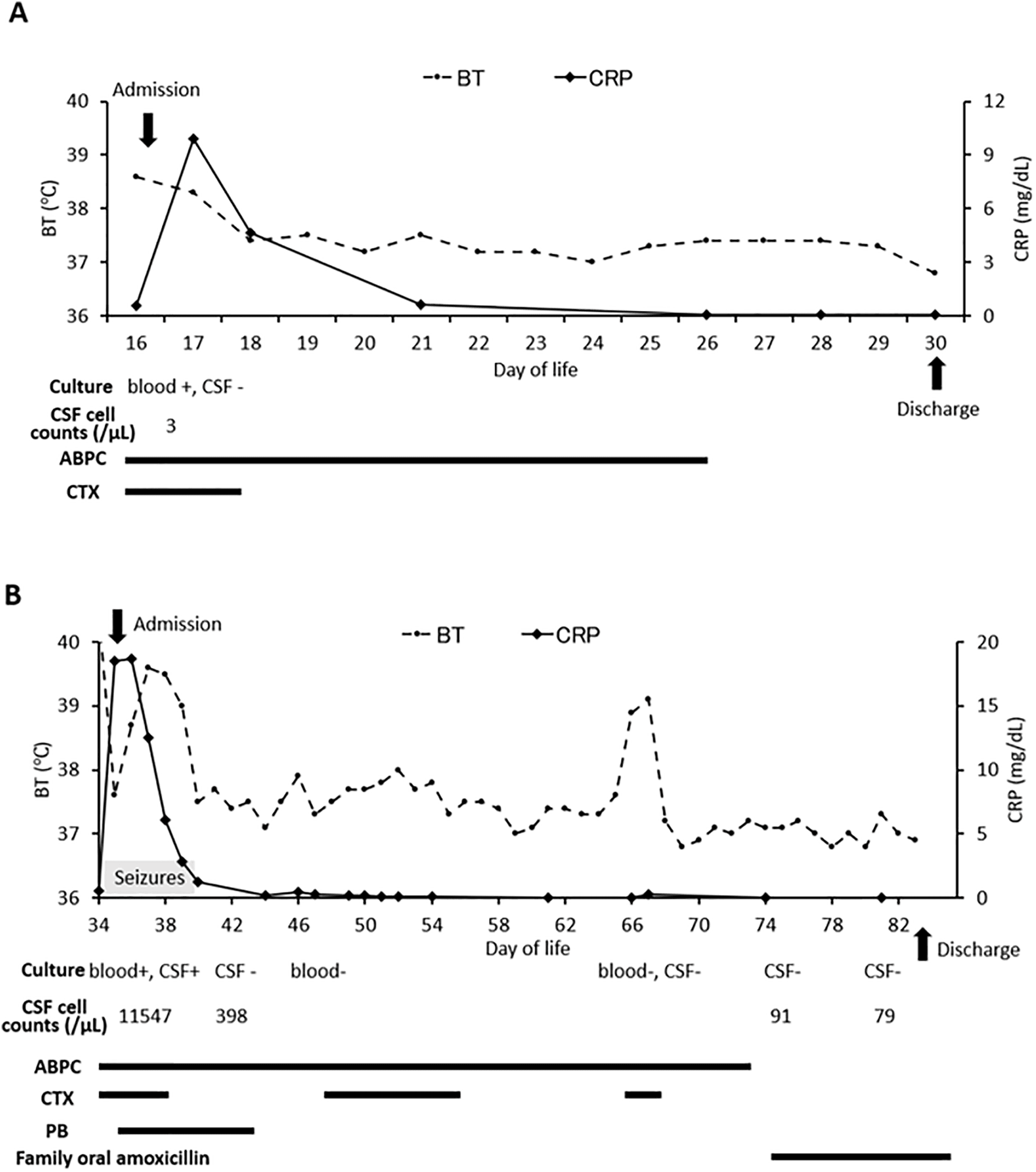
Figure 1. Clinical course of the patient. (A) First episode of hospitalization; (B) second episode of hospitalization. BT: body temperature; CRP: C-reactive protein; CSF: cerebrospinal fluid; ABPC: ampicillin; CTX: cefotaxime; PB: phenobarbital.
Four days after discharge, the infant again developed a fever of 39.5°C. On admission, she presented in a pre-shock state, with a heart rate of 198 beats/min, lethargy with pallor, a capillary refill time of 3 s, and a bulging anterior fontanelle. She received supportive treatment for shock and was started on intravenous ABPC and CTX. Both blood and CSF cultures yielded GBS this time. On the following day, she experienced three seizures, which were controlled with phenobarbital (Fig. 1B). An infection work-up, including contrast-enhanced magnetic resonance imaging (MRI) of the entire spine, contrast-enhanced brain MRI, and contrast-enhanced CT from the neck to the pelvis, revealed no significant abnormalities. Immunological studies demonstrated normal lymphocyte proliferative responses to phytohemagglutinin (stimulation index = 28.6) and concanavalin A (stimulation index = 44.7). Flow cytometric analysis revealed T cells 57.1%, B cells 23.0%, and natural killer cells 19.1%. CD4+ and CD8+ subsets accounted for 36.8% and 18.2%, respectively, with a CD4/CD8 ratio of 2.02. Serum IgG was 356 mg/dL. No abnormalities were detected, and no Toll-like receptor–related gene variants were identified.
GBS isolates from the blood cultures of both episodes and from the CSF culture of the second episode were analyzed for serotype and sequence type. All three isolates were identified as serotype III. Multilocus sequence typing revealed sequence type 17. Antimicrobial susceptibility testing showed susceptibility to penicillin G, ABPC, CTX, vancomycin, and ciprofloxacin, but resistance to erythromycin and clindamycin. One week after treatment initiation, the CSF culture became negative. The patient ultimately received a total of 39 days of intravenous ABPC therapy and remained hospitalized. After completion of antibacterial therapy, repeat CSF cultures remained negative, and the CRP levels stayed within the normal range.
Maternal cultures for GBS were negative in breast milk, throat, rectum, and vagina (tested twice). As transmission within the household was suspected, both parents and two siblings received a 10-day course of oral amoxicillin. Following this intervention, the patient was discharged in good condition, with no recurrence of invasive GBS disease. She received all immunizations according to the national schedule without any adverse reactions, and an adequate antibody response was confirmed after hepatitis B vaccination. Follow-up brain MRI at 6 and 18 months of age showed no abnormalities. The patient achieved normal developmental milestones, having rolled over at 5 months, sat without support at 8 months, walked independently at 11 months, and spoken her first words at 15 months.
Discussion
This case demonstrated recurrent late-onset invasive GBS infection that led a term infant to a life-threatening condition and highlighted the considerable medical and societal burden of recurrent GBS disease. Several studies have examined the risk factors for recurrent invasive GBS infection. A large cohort study from Japan identified prematurity and maternal colonization as significant contributors, while a recent French nationwide study similarly reported that preterm birth or very low birth weight was frequently associated with recurrence.4,6 Other proposed risk factors include multiple births, immunodeficiency, human immunodeficiency virus exposure, and insufficient duration of antibiotic therapy.7,8 However, our patient exhibited none of these conditions, suggesting that even healthy term infants may be vulnerable to severe recurrent invasive GBS disease.
GBS serotype III and clonal complex 17 have been reported significantly more frequently in recurrent cases, consistent with our patient’s presentation.4,9,10 Empirical antibiotic therapy generally includes amoxicillin or CTX in combination with amikacin or gentamicin, administered for approximately 10 days in accordance with current recommendations.11 Although clonal complex 17 has been suggested to be hypervirulent and associated with LOD and recurrence, its antimicrobial susceptibility is not different from that of other sequence types, and a recent retrospective observational study reported no β-lactam–tolerant strains, supporting the effectiveness of standard antibiotic therapy.4 In many infants, recurrence is thought to result from reinvasion of persistently colonized mucosal sites or re-exposure to a household carrier. Reports indicate that breast milk accounts for nearly half of recurrent GBS disease cases, and in some infants, sibling-to-sibling transmission has been documented. Furthermore, studies have shown that recurrence often involves the same strain as the initial infection, supporting the hypothesis of persistent colonization rather than reinfection with a new strain.5,12 However, data on family colonization sources are often insufficient, making precise identification of the pathogen source difficult. Similar to our case, although maternal breast milk and body sites were screened and found to be negative for GBS, transmission from another family member or transient colonization could not be excluded.
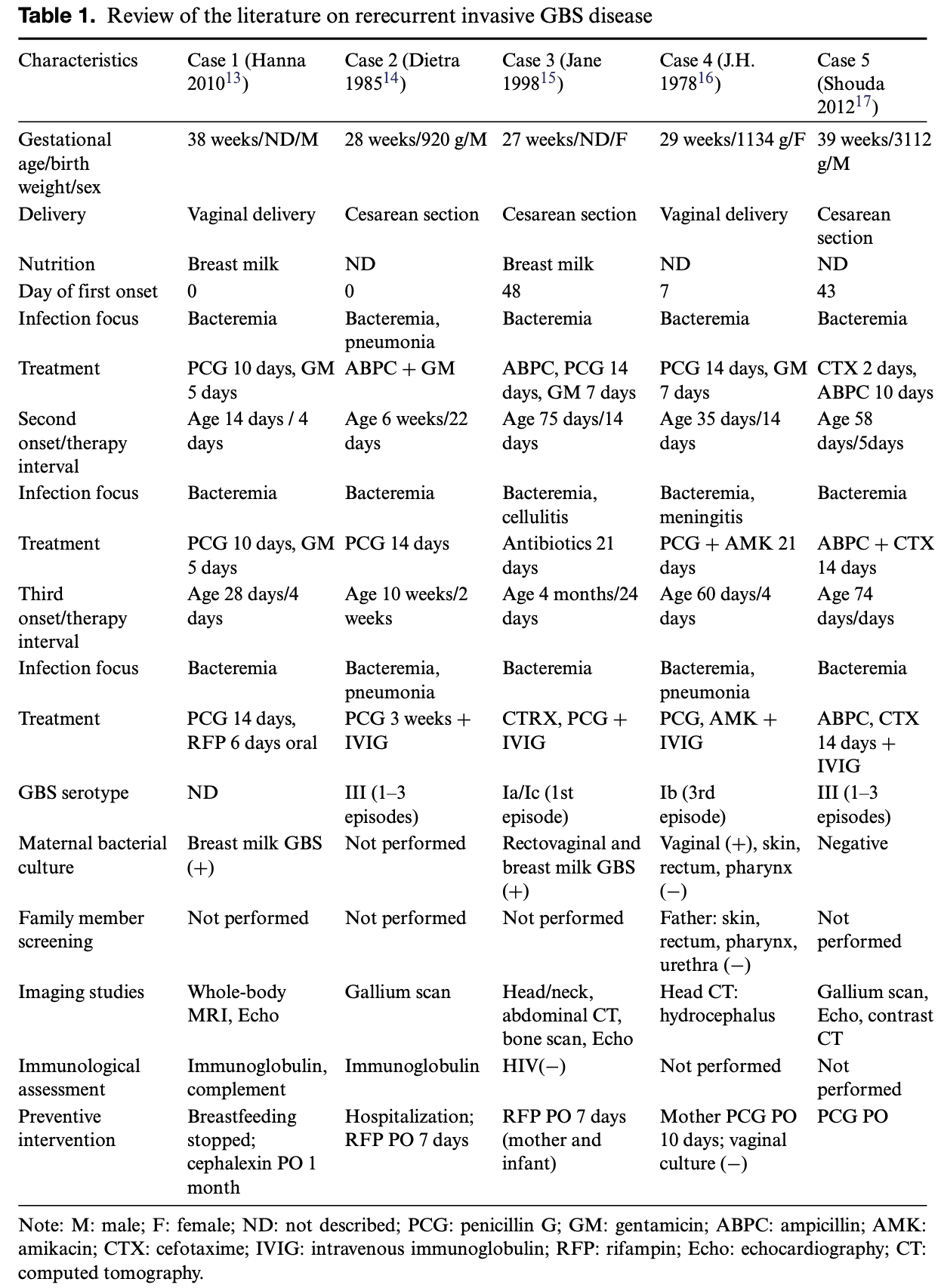
Therefore, preventive strategies against recurrence are necessary, even in term and immunocompetent infants. There are few studies focusing on the prevention of recurrent GBS disease. To gather insights into possible approaches, we reviewed case reports relevant to rerecurrent GBS disease and assessed the preventive measures described (Table 1).13–17 Initial steps often include immunological and imaging evaluations to exclude underlying abnormalities that might predispose to reinfection. Maternal bacterial screening is frequently performed to help identify the source of infection. Considering that breast milk has been reported as a frequent source of late-onset infection, interruption of breastfeeding or pasteurization may be effective, as illustrated in Case 1. When GBS is detected in maternal cultures, oral antibiotic therapy for the mother has also been suggested, as in Case 4. Prolonged or repeated antibiotic therapy for the infant after hospitalization may also contribute to preventing recurrence (Cases 2, 3, and 5). Although paternal cultures were examined only in Case 4 and were negative, the possibility of household transmission should not be overlooked. In our case, although we were unable to perform comprehensive family cultures, prophylactic oral antibiotics administered to both parents and siblings successfully prevented further recurrence. This emphasizes the potential role of household-targeted interventions, although ideally GBS screening should be performed prior to prophylaxis. In addition, as observed in Case 2 and in our patient, extended hospitalization after completion of antibiotic therapy allowed for close monitoring and temporary separation from potential household sources of infection. Furthermore, intravenous immunoglobulin (IVIG) administration has been reported to increase anti-GBS type III antibody levels and may serve as immunoprophylaxis against horizontal transmission in very low birth weight infants. This approach could also be considered in recurrent cases.18
There are several limitations to this case report. Although horizontal transmission was suspected, institutional regulations prevented GBS screening of other family members, including the father and two siblings, making it impossible to confirm the source of the GBS isolates. In addition, the single-case nature of this report limits the generalizability of the proposed preventive strategies. Nevertheless, the optimal preventive strategies remain uncertain, and evidence-based guidelines are lacking. Large-scale studies are required to identify the most effective approaches. Until maternal GBS vaccines become widely available, heightened surveillance, early recognition of recurrence, and consideration of preventive measures remain essential in clinical practice.19
Conclusion
This case highlights that recurrent invasive GBS infection can occur even in term, immunocompetent infants. Although infectious colonization was not detected by maternal screening, prolonged hospitalization for close monitoring and prophylactic oral antibiotic therapy for family members successfully prevented further recurrence.
Acknowledgment
We would like to extend our gratitude for the patient’s cooperation in the data collection.
Funding Source
No financial support was provided.
Author Contributions
M.N. and Y.Z. drafted the original manuscript. M.N., Y.S., and Y.Z. were responsible for data curation and interpretation. H.T., S.O., A.S., T.M., M.E., M.N., T.E. and T.T. substantially contributed to the manuscript draft. All authors have read and approved the manuscript and agree with its content and data.
Data Availability Statement
The datasets used in this study are available from the corresponding author upon reasonable request.
Ethical Statement
The article does not involve the participation of any animals. The patient has provided written informed consent for the publication of this report and the accompanying images.
Conflict of Interest
The authors report no conflicts of interest in this work.
References
[1] Hanna M, Noor A. Streptococcus Group B. StatPearls; 2025.
[2] Coggins SA, Puopolo KM. Neonatal group B streptococcus disease. Pediatr Rev. February 1, 2024;45(2):63–73. doi:10.1542/pir.2023-006154.
[3] Miselli F, Frabboni I, Di Martino M, et al. Transmission of Group B Streptococcus in late-onset neonatal disease: a narrative review of current evidence. Ther Adv Infect Dis. January–December 2022;9:20499361221142732. doi:10.1177/20499361221142732.
[4] Sbaa G, Delettre N, Guyonnet C, et al. Recurrent group B streptococcus neonatal invasive infections, France, 2007–2021. J Infect Dis. February 20, 2025;231(2):329–333. doi:10.1093/infdis/jiae307.
[5] Freudenhammer M, Karampatsas K, Le Doare K, et al. Invasive group B streptococcus disease with recurrence and in multiples: towards a better understanding of GBS late-onset sepsis. Front Immunol. 2021;12:617925. doi:10.3389/fimmu.2021.617925.
[6] Shibata M, Matsubara K, Matsunami K, et al. Epidemiology of group B streptococcal disease in infants younger than 1 year in Japan: a nationwide surveillance study 2016–2020. Eur J Clin Microbiol Infect Dis. April 2022;41(4):559–571. doi:10.1007/s10096-021-04396-y.
[7] Moon CJ, Kwon TH, Lee KS, et al. Recurrent neonatal sepsis and progressive white matter injury in a premature newborn culture-positive for group B Streptococcus: a case report. Medicine (Baltimore). June 25, 2021;100(25):e26387. doi:10.1097/MD.0000000000026387.
[8] Suresh S, Tyrrell G, Alhhazmi A, Escoredo S, Hawkes M. Recurrent sepsis and neuroinvasive disease in a neonate culture-positive for a group B streptococcus CPS III serotype, hvgA+ strain. JMM Case Rep. June 2016;3(3):e005034. doi:10.1099/jmmcr.0.005034.
[9] Zhou Y, Wang LQ, Yan Q, et al. Genomic analysis of group B streptococcus from neonatal sepsis reveals clonal CC17 expansion and virulence- and resistance-associated traits after intrapartum antibiotic prophylaxis. Clin Infect Dis. December 19, 2022;75(12):2153–60. doi:10.1093/cid/ciac331.
[10] Tazi A, Plainvert C, Anselem O, et al. Risk factors for infant colonization by hypervirulent CC17 group B streptococcus: toward the understanding of late-onset disease. Clin Infect Dis. October 30, 2019;69(10):1740–1748. doi:10.1093/cid/ciz033.
[11] Puopolo KM, Lynfield R, Cummings JJ, Committee On F, Newborn, Committee On Infectious D. Management of infants at risk for group B streptococcal disease. Pediatrics. August 2019;144(2):1. doi:10.1542/peds.2019-1881.
[12] Green PA, Singh KV, Murray BE, Baker CJ. Recurrent group B streptococcal infections in infants: clinical and microbiologic aspects. J Pediatr. December 1994;125(6 Pt 1):931–938. doi:10.1016/s0022-3476(05)82012-8.
[13] Soukka H, Rantakokko-Jalava K, Vahakuopus S, Ruuskanen O. Three distinct episodes of GBS septicemia in a healthy newborn during the first month of life. Eur J Pediatr. October 2010;169(10):1275–1277. doi:10.1007/s00431-010-1187-6.
[14] Millard DD, Bussey ME, Shulman ST, Yogev R. Multiple group B streptococcal infections in a premature infant: eradication of nasal colonization with rifampin. Am J Dis Child. October 1985;139(10):964–965. doi:10.1001/archpedi.1985.02140120010009.
[15] Atkins JT, Heresi GP, Coque TM, Baker CJ. Recurrent group B streptococcal disease in infants: who should receive rifampin? J Pediatr. March 1998;132(3 Pt 1):537–539. doi:10.1016/s0022-3476(98)70037-x.
[16] McCrory JH, Au-Yeung YB, Sugg VM, Chiu TT, Garrison RD. Recurrent group B streptococcal infection in an infant: ventriculitis complicating type Ib meningitis. J Pediatr. February 1978;92(2):231–233. doi:10.1016/s0022-3476(78)80011-0.
[17] Shoda T, Miyagi N, Shirai K, et al. Double recurrence of group B streptococcus bacteremia in an immunocompetent infant. Pediatr Int. October 2012;54(5):701–703. doi:10.1111/j.1442-200X.2011.03492.x.
[18] Takayanagi T, Tanaka H, Yoshinaga M, et al. An outbreak of group B streptococcus infection in a neonatal nursery and subsequent trial for prophylaxis of nosocomial transmission. Acta Paediatr Jpn. February 1994;36(1):88–90. doi:10.1111/j.1442-200x.1994.tb03137.x.
[19] Joel B, Moses V, Veeraraghavan B, et al. Group B streptococcus vaccine update: a crucial public health initiative for India. Indian J Med Microbiol. July 11, 2025;57:100917. doi:10.1016/j.ijmmb.2025.100917.
 |
Copyright: © 2025 Nakamura et al. This work is licensed under a Creative Commons Attribution 4.0 International License, which permits unrestricted use, distribution, and reproduction in any medium, provided the original work is properly cited. |