| Journal of Clinical Question. 2026; 3(2): e113 https://doi.org/10.69854/jcq.2026.0012 Advance access publication date 25 April 2026 |
 |
Review
Prevention and Therapeutic Development in Mitochondrial Disease: A Review of Recent Real-World Clinical Evidence

1Department of Pediatrics, Faculty of Medicine, Saga University, Saga, Japan.
2Department of Pediatrics, Hokuto Hospital, Hokkaido, Japan.
*Corresponding Author: e-mail: yumeng1990228@gmail.com
Submitted: February 17, 2026 Accepted: April 24, 2026
Clinical Question Box
How is clinical research in primary mitochondrial disease progressing?
Recent years have seen more rigorous studies of primary mitochondrial disease. Genotype-directed therapies have improved outcomes in selected disorders, and gene therapy for Leber hereditary optic neuropathy has demonstrated sustained visual benefits. Preventive strategies have also advanced, including preimplantation genetic testing for monogenic diseases and the first regulated clinical application of mitochondrial donation in carefully selected patients.
Abstract
Although individually rare, primary mitochondrial diseases (PMD) collectively represent a substantial cause of inherited metabolic and multisystem diseases. Advances in genomic testing have broadened the recognized spectrum of mitochondrial diseases and improved molecular diagnosis, while progress in disease modeling has accelerated therapeutic development. Concurrently, an expanding body of clinical research has evaluated supportive, pharmacological, preventive, and emerging advanced interventions across genetically and clinically heterogeneous mitochondrial disease subtypes. However, the available evidence remains fragmented, with considerable variability in study design, patient selection, outcome measures, and follow-up duration. In this review, we summarize recent therapeutic and preventive interventions for PMD, including supplement and dietary strategies, mechanism-based drug therapies, reproductive prevention approaches for mitochondrial DNA disease, and emerging advanced therapies such as gene, cell, and enzyme-based treatments. We also examine the strengths and limitations of the current evidence base and discuss implications for clinical practice, trial design, and future translational research.
Keywords: Mitochondrial diseases, clinical trials, reproductive prevention, mitochondrial donation
Introduction
Mitochondria are semi-autonomous organelles present in multiple copies in most human cells. They are essential for cellular energy metabolism through the mitochondrial respiratory chain and oxidative phosphorylation (OXPHOS), which generate adenosine triphosphate (ATP). Mitochondrial function relies on proteins encoded by both mitochondrial DNA (mtDNA) and nuclear DNA. Pathogenic variants in either genome can lead to primary mitochondrial diseases (PMDs)—a genetically heterogeneous group of inherited metabolic disorders.1 These disorders affect approximately 1 in 4,300 adults and 1 in 5,000 live births, often presenting with progressive, multisystem involvement.2,3
The pathophysiology of PMDs extends beyond ATP deficiency to encompass oxidative stress, impaired mitochondrial quality control, and signaling abnormalities. PMDs primarily affect high-energy-demand organs such as the brain, skeletal muscle, heart, liver, and kidneys. In mtDNA-related disorders, heteroplasmy and tissue-specific biochemical thresholds further shape the clinical expression of PMDs, leading to wide variation in age at onset, severity, and organ involvement.4 The clinical spectrum includes Leigh syndrome, mitochondrial encephalomyopathy, lactic acidosis, and stroke-like episodes (MELAS), and Leber hereditary optic neuropathy (LHON), as well as cardiomyopathies and skeletal myopathies. This heterogeneity complicates diagnosis, outcome assessment, and therapeutic development.
Most PMDs lack curative therapy, with limited disease-modifying treatment options. Current management remains largely supportive, focusing on symptom control, rehabilitation, nutritional optimization, and prevention of metabolic decompensation. Supplements such as coenzyme Q10 (CoQ10) and riboflavin, as well as dietary strategies such as the ketogenic diet in selected settings, are widely used, though the strength of evidence varies across interventions and disease subtypes. Advances in next-generation sequencing have improved molecular diagnosis and patient stratification, facilitating more targeted clinical studies and therapeutic development.5 At the same time, the therapeutic landscape is expanding beyond empirical supportive care to include mechanism-based pharmacological agents, reproductive prevention strategies for mtDNA disease, and advanced approaches such as gene, cell, and enzyme-based therapies.6 However, the available evidence remains fragmented, with substantial heterogeneity in study design, patient selection, outcome measures, and duration of follow-up.
This review provides a clinically oriented synthesis of real-world clinical evidence in PMD, spanning supportive, pharmacological, preventive, and emerging advanced therapeutic strategies. The studies discussed were selected for their clinical relevance, recency, and contribution to illustrating the current direction of therapeutic development in genetically and phenotypically heterogeneous mitochondrial disorders. We summarize therapeutic and preventive interventions in PMDs with particular attention to translational progress, implications for future trial design and patient care, and the extent to which current clinical applicability is relatively established or remains preliminary.
Supplement and Dietary Interventions
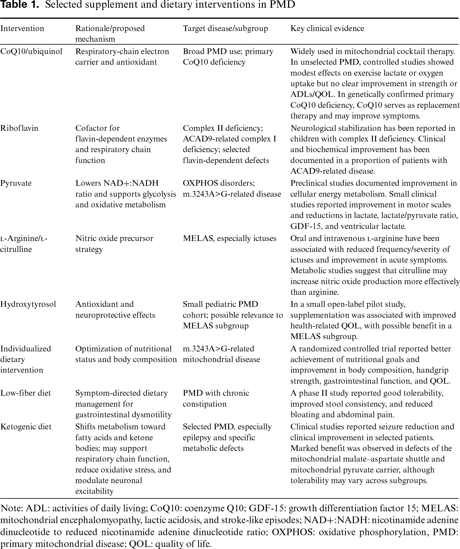
Supplement-based and dietary approaches remain central to PMD supportive care, encompassing empirical vitamin and cofactor supplementation, metabolically targeted compounds, individualized nutritional support, and dietary therapies such as the ketogenic diet (Table 1).
Empiric Supplements and Mitochondrial Cocktails
Empirical supplement regimens for PMDs often include CoQ10, riboflavin, l-carnitine, thiamine, biotin, alpha-lipoic acid, niacin, and other vitamins or cofactors, although supporting evidence varies considerably among individual agents and prescribing practices differ across centers.7,8 Among these agents, CoQ10 has been the most extensively studied. As an electron carrier in the respiratory chain and an antioxidant, CoQ10 remains a core component of mitochondrial cocktail regimens, though evidence for its benefit in unselected mitochondrial disease is limited. In an early study of 16 clinically heterogeneous patients treated with CoQ10 in combination with multiple vitamins, plasma CoQ10 levels increased substantially, while objective metabolic indices and overall clinical outcomes showed no improvement.9 A randomized, double-blind, crossover trial of 30 patients receiving 1,200 mg/day for 60 days reported attenuation of post-exercise lactate rise and a modest increase in early exercise oxygen uptake, but no clear improvement in muscle strength, resting lactate, or quality of life.10 In contrast, in genetically confirmed primary CoQ10 deficiency, CoQ10 serves as a replacement therapy and may improve symptoms in some patients.11 Recent reviews also note that, despite its widespread use, high-quality clinical evidence supporting CoQ10 supplementation remains limited, particularly in children and adolescents.12
Riboflavin is another commonly used component of empirical supplement therapy, with a more clearly defined role in selected disorders. In children with complex II deficiency, riboflavin treatment was reported to stabilize neurological status in a small case series of three patients.13 Riboflavin has also been used in ACAD9-related complex I deficiency, where clinical and biochemical improvement has been reported in a substantial proportion of patients. More broadly, riboflavin is often employed in conditions involving flavin-dependent respiratory chain dysfunction, although the strength of evidence varies according to the underlying genotype and phenotype.14
Metabolic and Redox-Targeted Supplementation
Several nutritional and metabolic interventions have been developed to improve cellular redox balance and lactate metabolism. Pyruvate therapy aims to reduce the elevated nicotinamide adenine dinucleotide to reduced nicotinamide adenine dinucleotide (NAD+:NADH) ratio associated with OXPHOS dysfunction and thereby facilitate glycolytic flux and oxidative metabolism. In experimental work, pyruvate exerted marked and sustained effects on energy metabolism in cybrid cells carrying the m.3243A>G mutation.15 Clinically, among four bedridden pediatric patients with OXPHOS disorders treated with sodium pyruvate at 0.5–1.0 g/kg/day for more than 12 months, three showed improvement in Newcastle Pediatric Mitochondrial Disease Scale (NPMDS) and Gross Motor Function Measure-88 scores.16 In a 48-week prospective exploratory study of 11 Japanese adults with genetically, biochemically, and clinically confirmed mitochondrial disease, pyruvate therapy was associated with significant reductions in plasma lactate, lactate/pyruvate ratio, serum growth differentiation factor 15 (GDF-15), and lateral ventricular lactate, alongside a decreasing trend in total Newcastle Mitochondrial Disease Adult Scale (NMDAS) score.17
l-Arginine has been studied most extensively in MELAS, particularly in relation to stroke-like episodes or ictuses. MELAS is a progressive and potentially life-threatening mitochondrial encephalomyopathy characterized by recurrent ictuses.18 In a clinical trial of oral l-arginine in patients with the m.3243A>G mtDNA mutation who had experienced ictuses within the past 2 years, treatment at 0.3–0.5 g/kg/day for 2 years significantly prolonged the interictal period and reduced the incidence and severity of ictuses.19 Intravenous l-arginine has also been evaluated in acute settings, administered at 0.5 g/kg per dose to patients presenting within six hours of ictus onset, with a further 0.5 g/kg added if symptoms persisted. This regimen was associated with reductions in headache, nausea/vomiting, impaired consciousness, and visual disturbance.19 l-Citrulline has also attracted interest as a nitric oxide precursor in MELAS, with metabolic studies suggesting that it may enhance nitric oxide production more effectively than arginine.20
Hydroxytyrosol, a phenolic compound with antioxidant properties, has also been investigated. Preclinical studies have suggested neuroprotective effects against oxidative stress and neurodegeneration.21 In a small open-label pilot study involving nine pediatric patients with mitochondrial disease, hydroxytyrosol supplementation was associated with improved health-related quality of life as measured by the Pediatric Quality of Life Inventory, with possible benefit in a subgroup of patients with MELAS.22
Nutritional Support and Dietary Intervention
Beyond individual supplements, nutritional status is increasingly recognized as an important modifier of disease burden in mitochondrial disease. Malnutrition is common in the pediatric population and may exacerbate symptoms by further compromising mitochondrial function.23,24 In patients with m.3243A>G-related mitochondrial disease, a randomized controlled trial of an individually tailored dietary intervention showed that the intervention group more frequently achieved prespecified nutritional goals and demonstrated improvements in body composition, handgrip strength, gastrointestinal function, and quality of life compared with controls.25
Consistent with the high prevalence of gastrointestinal dysmotility in mitochondrial disease, a phase II study of a low-fiber diet (<10 g/day) reported good tolerability, along with improved stool consistency and reduced gastrointestinal symptoms, including bloating and abdominal pain, in patients with chronic constipation.26
Ketogenic Diet
The ketogenic diet has received increasing attention as a dietary therapy for selected mitochondrial disorders. By redirecting energy utilization from carbohydrates toward fatty acids and ketone bodies, this diet mimics fasting and may support mitochondrial metabolism through several mechanisms, including enhancement of respiratory chain energy production, limitation of glycolytic flux, increased fatty acid oxidation, reduction of oxidative stress, and modulation of neuronal excitability.27,28
Additional proposed mechanisms include metabolic gene reprogramming, enhanced mitochondrial biogenesis, and regulation of seizure pathways via peroxisome proliferator–activated receptor-γ and astrocyte metabolism.28 Clinical studies suggest that the ketogenic diet may be particularly useful in patients with epilepsy and selected metabolic subtypes. In 2022, Huang et al. reported that 1- and 3-month ketogenic diet interventions achieved >50% seizure reduction in 31.8–72.3% of patients with mitochondrial disease.29 In another series, clinical improvement was observed in 9 of 11 patients aged 1 month to 18 years treated with the ketogenic diet, with outcomes appearing more favorable than in patients who did not receive the diet.30 The ketogenic diet has also been reported to be highly effective in defects of the mitochondrial malate–aspartate shuttle and the mitochondrial pyruvate carrier, with benefit observed in 11 of 13 patients, particularly with marked seizure reduction.31 However, more recent studies indicate that tolerability and safety may vary across mitochondrial disease subgroups, and modified ketogenic approaches may be unsuitable for patients with mtDNA deletion-related myopathy.32 Accordingly, the ketogenic diet should be viewed as a selective rather than universal dietary strategy in mitochondrial disease.
Overall, supplement and dietary interventions continue to play an important role in supportive care for PMD, but their clinical relevance is not uniform across disorders. The strongest practical indications are generally confined to selected settings, such as replacement therapy in primary CoQ10 deficiency, riboflavin-responsive defects, l-arginine-based management in MELAS, and the ketogenic diet in selected epilepsy-predominant or metabolic phenotypes. For many empiric supplements used across broad PMD populations, however, the available evidence remains limited, heterogeneous, and insufficient to support generalized disease-modifying benefits.
Drug Therapy
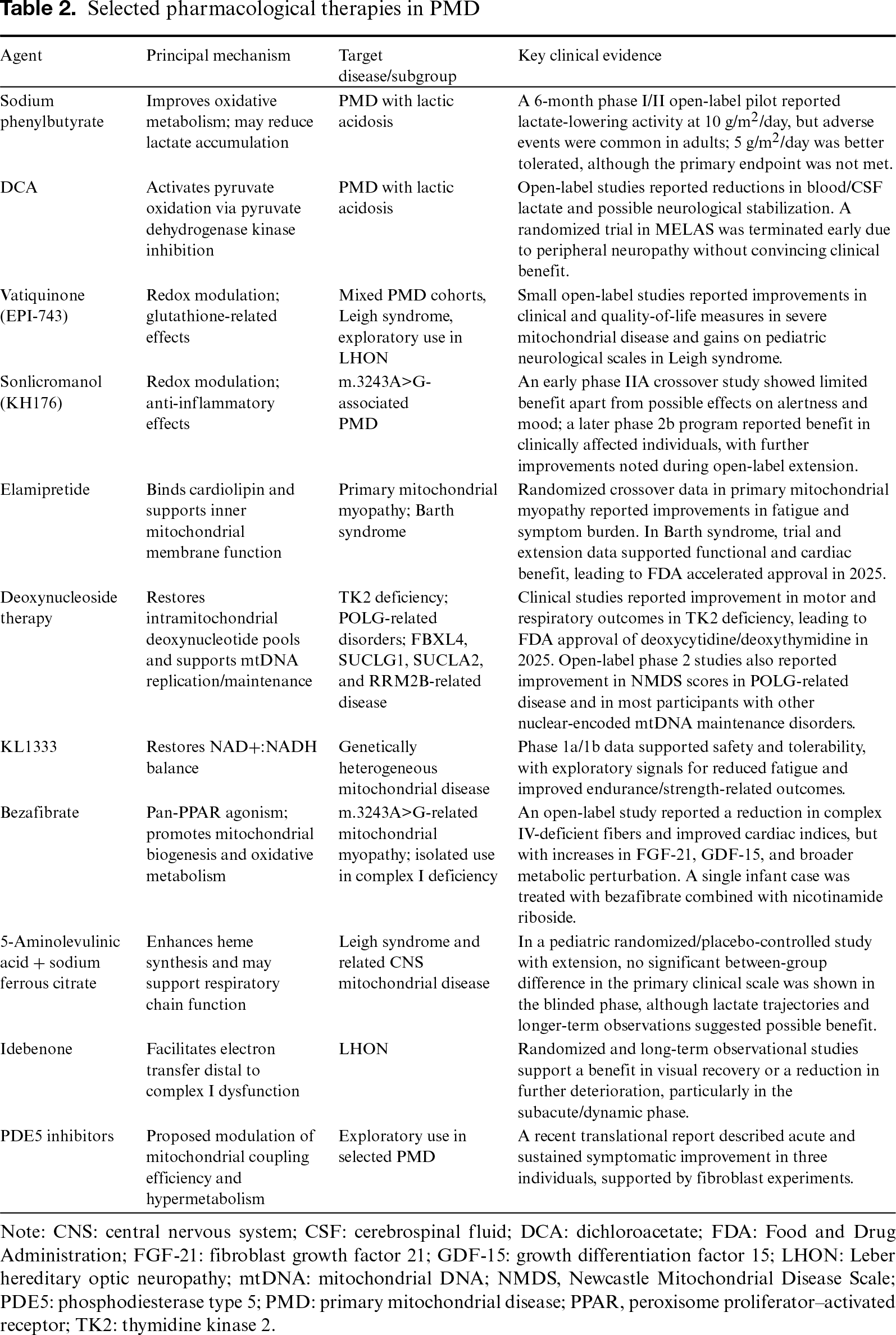
Pharmacological treatment of PMD is shifting from empirical metabolic support toward mechanism-based intervention. A growing number of agents targeting key pathogenic pathways—including pyruvate metabolism, redox regulation, cardiolipin remodeling, mtDNA maintenance, NAD(H) homeostasis, and respiratory chain dysfunction—have entered clinical development. At the same time, therapeutic translation remains difficult owing to marked genetic and phenotypic heterogeneity, small and fragmented study populations, and the challenge of translating biochemical effects into consistent clinical benefits (Table 2).
Agents Targeting Pyruvate Metabolism and Lactic Acidosis
Sodium phenylbutyrate has attracted renewed interest as a treatment for mitochondrial lactic acidosis based on preclinical evidence indicating improved mitochondrial function and oxidative capacity.33 In a recent 6-month phase I/II open-label pilot study in patients with PMD and lactic acidosis, treatment at 10 g/m2/day reduced lactate exposure, though adverse events were common in adults. A lower dose of 5 g/m2/day was better tolerated but failed to meet the primary endpoint.34 Although these data indicate biological activity, dose-limiting toxicity and uncertain clinical efficacy remain key limitations.
Dichloroacetate (DCA), a pyruvate dehydrogenase kinase inhibitor that promotes pyruvate oxidation, has long been investigated as a lactate-lowering agent in mitochondrial disorders associated with lactic acidosis.35 Early open-label studies reported reductions in blood and cerebrospinal fluid lactate, alongside possible stabilization of neurological symptoms.36 Subsequent controlled studies—particularly in MELAS—highlighted peripheral neurotoxicity as a major safety concern and failed to demonstrate consistent clinical benefits despite measurable effects on lactate metabolism.37 The therapeutic role of DCA in PMDs therefore remains limited.
Redox-Modulating Therapies
Redox imbalance is a recurrent feature of mitochondrial disease that is increasingly recognized as an important therapeutic target. Vatiquinone (EPI-743), a para-benzoquinone derivative developed as a redox-active analogue of CoQ10, was designed to overcome the pharmacological limitations of CoQ10 and idebenone. Early open-label studies reported signals of benefit across several mitochondrial phenotypes. In a subject-controlled emergency treatment study involving patients with severe mitochondrial disease at high risk of near-term clinical deterioration, most survivors showed improvement in clinical and quality-of-life measures, with imaging findings correlating with clinical response.38 In children with genetically confirmed Leigh syndrome, a phase 2A open-label trial reported improvement in motor and disease severity measures, alongside altered glutathione homeostasis.39,40 A small exploratory study in acute LHON also reported possible benefits in visual outcomes.41 These studies suggest potential clinical and biochemical activity of vatiquinone in selected mitochondrial phenotypes, although interpretation remains limited by small sample size, open-label design, and the lack of controlled confirmation.
Sonlicromanol (KH176) is another redox-modulating compound under clinical development. Its proposed mechanism includes restoration of redox balance and anti-inflammatory effects mediated by inhibition of microsomal prostaglandin E synthase-1.42,43 A phase 2b development program on adults with m.3243A>G-associated PMD reported benefits across several domains, with additional changes observed during the open-label extension phase.44 By contrast, an earlier crossover phase IIA study did not demonstrate significant improvement in gait or most secondary outcomes, apart from possible effects on alertness and mood.45 Findings to date have varied across studies.
Cardiolipin-Targeted Therapy
Elamipretide is a mitochondria-targeted tetrapeptide that binds cardiolipin in the inner mitochondrial membrane and has been shown to improve membrane stability, respiratory efficiency, and ATP production.46 A randomized, double-blind, placebo-controlled crossover trial in primary mitochondrial myopathy demonstrated that short-course daily subcutaneous elamipretide alleviated patient-reported fatigue and muscle symptoms, with a clinically meaningful improvement in the 6-minute walk test.47 The most substantial clinical outcome was seen in a 28-week randomized placebo-controlled trial and a 168-week open-label extension on Barth syndrome, which showed sustained tolerability alongside improvement in functional assessments and cardiac function.48 Additional case-series data suggest that weight-based dosing is well tolerated in pediatric patients.49 Based on these trial and extension data, elamipretide received accelerated approval in the United States in 2025 as the first therapy for Barth syndrome. Outside Barth syndrome, however, broader applicability across heterogeneous PMD populations remains less certain.
Nucleoside Therapy for mtDNA Maintenance Disorders
Nucleoside supplementation has emerged as one of the clearest examples of genotype-directed therapy in mitochondrial medicine. In thymidine kinase 2 (TK2) deficiency, treatment with deoxycytidine and deoxythymidine is intended to restore intramitochondrial nucleotide pools and improve mtDNA replication. Clinical studies have demonstrated improvements in motor and respiratory outcomes, and this strategy has now been translated into regulatory approval. The success of nucleoside therapy in TK2 deficiency provides proof-of-principle that targeted correction of nucleotide imbalance can modify the course of mtDNA maintenance disorders.50,51
This approach is now being evaluated in other disorders of mtDNA maintenance. In an open-label phase 2 study, patients with POLG-related disease exhibited improvement in Newcastle Mitochondrial Disease Scale scores, with stable or reduced GDF-15 levels during treatment.52 Similar observations were reported in a phase 2 study of patients with pathogenic variants in FBXL4, SUCLG1, SUCLA2, or RRM2B.53 Although these studies were limited and uncontrolled, they suggest that manipulation of nucleotide homeostasis may have broader applicability beyond TK2 deficiency.
NADH Modulation and Metabolic Reprogramming
KL1333 is an orally administered small molecule designed to restore the intracellular NAD+:NADH ratio, thereby supporting OXPHOS and ATP generation.54 In a phase 1a/1b study conducted on adults with mitochondrial disease, KL1333 was generally well tolerated, with gastrointestinal adverse events indicating dose dependence. Exploratory analyses suggested a possible reduction in fatigue and improvement in measures of functional strength and endurance.55 Although these findings are preliminary, KL1333 is of interest because it targets a central metabolic disturbance that may be relevant across multiple mitochondrial disease phenotypes.
Bezafibrate, a pan-peroxisome proliferator-activated receptor agonist that promotes mitochondrial biogenesis and oxidative metabolism, has also been investigated as a metabolic therapy. In an open-label study of adults with m.3243A>G-related mitochondrial myopathy, bezafibrate reduced the proportion of complex IV-deficient muscle fibers and improved cardiac parameters. However, these changes were accompanied by increases in circulating biomarkers such as fibroblast growth factor 21 and GDF-15, as well as broader metabolic disturbances, raising concerns regarding long-term tolerability.56 Isolated case reports suggest potential benefit of bezafibrate combined with nicotinamide riboside in infantile complex I deficiency.57 Further studies are needed to define the clinical role of bezafibrate in mitochondrial disease.
Respiratory Chain Support and Heme Pathway Augmentation
5-Aminolevulinic acid combined with sodium ferrous citrate has been shown to enhance heme production and upregulate mitochondrial respiratory chain activity in vitro and in vivo.58 In a Japanese clinical study comprising a double-blind, placebo-controlled phase, an open-label phase, and subsequent long-term administration in pediatric patients with Leigh syndrome and central nervous system disorders, sustained treatment was associated with a gradual reduction in average serum lactate levels and stabilization or improvement in NPMDS scores.59
Genotype-Specific Therapy in LHON
Idebenone remains the most established pharmacological therapy for LHON. As a short-chain benzoquinone, idebenone can facilitate electron transfer downstream of complex I dysfunction and may help preserve or reactivate viable retinal ganglion cells. Evidence from a randomized, placebo-controlled trial, together with subsequent follow-up and observational studies, supports beneficial effects on visual outcomes, including reduced risk of further visual decline and promotion of visual recovery across different stages of the disease.60–62 These findings support idebenone as an established treatment option for LHON.
Emerging Repurposing Strategies
Drug repurposing has recently attracted increasing interest in mitochondrial medicine. A recent translational study suggested that phosphodiesterase type 5 inhibitors may improve mitochondrial coupling efficiency and reduce hypermetabolism. In that report, tadalafil treatment was associated with acute and sustained symptomatic improvement in three individuals with PMD, supported by complementary fibroblast data.63 These observations are preliminary and warrant further evaluation.
Taken together, the current pharmacological landscape in PMD is best interpreted in terms of evidentiary maturity and disease specificity. The clearest clinically actionable signals are currently seen in selected genotype- or disease-specific contexts, such as idebenone in LHON and deoxynucleoside therapy in TK2 deficiency. By contrast, many other agents targeting redox balance, cardiolipin, lactate metabolism, or NAD(H) homeostasis remain promising but investigational, with interpretation limited by small sample sizes, mixed cohorts, short follow-up, and non-comparative study designs.
Emerging Advanced Therapies
Although advanced therapeutic development in mitochondrial disease remains limited, several strategies have progressed beyond symptomatic treatment and aim to correct the primary molecular defect or modify downstream tissue injury. To date, the strongest clinical outcome has been observed in LHON, where gene replacement has emerged as the leading translational strategy. Most studies have focused on allotopic expression of the wild-type ND4 gene delivered via intravitreal rAAV2. Across the major ND4 programs, including early-phase and later confirmatory studies, unilateral treatment has demonstrated an acceptable safety profile, alongside clinically meaningful improvements in best-corrected visual acuity that may persist for years after a single injection. A striking and consistently reported finding is improvement in the contralateral untreated eye, suggesting a bilateral biological effect despite unilateral administration, potentially related to vector or transgene transfer along the visual pathway.64,65 More recently, gene therapy has been extended to the rarer ND1 form of LHON. In a 2025 pilot study, unilateral intravitreal rAAV2-ND1 was well tolerated, and some patients showed improvement not only in the injected eye but also in binocular visual function, supporting the broader applicability of mutation-specific ocular gene replacement in mtDNA disease.66 Beyond LHON, however, gene therapy remains largely preclinical. In Barth syndrome, an X-linked disorder caused by TAZ mutations, systemic AAV9-mediated TAZ gene replacement restored mitochondrial structure, improved respiration, and ameliorated both cardiac and skeletal muscle dysfunction in animal models. These findings provide a strong proof-of-concept for nuclear gene replacement in mitochondrial disorders caused by defects in nuclear-encoded mitochondrial proteins, although clinical translation has not yet been achieved.67
Cell- and enzyme-based strategies have also been explored, though clinical evidence remains extremely limited. In LHON, the Stem Cell Ophthalmology Treatment Study investigated autologous bone marrow–derived stem cells delivered via combinations of retrobulbar, intravitreal, subtenon, intra-optic nerve, subretinal, and intravenous routes. The proposed mechanism included paracrine neuroprotection and possible transfer of functional mitochondria from donor cells to injured retinal ganglion cells. Reported visual gains in some patients were encouraging. However, since the study was uncontrolled and heterogeneous in both delivery route and disease severity, its findings should be interpreted cautiously.68 In contrast, a more mechanism-based metabolic replacement approach has shown promise in mitochondrial neurogastrointestinal encephalomyopathy. Erythrocyte-encapsulated thymidine phosphorylase, designed to reduce the toxic accumulation of thymidine and deoxyuridine in patients with TP deficiency, was associated with biochemical improvement in two of three treated patients, while weight gain and broader clinical improvement were observed in a subset of patients; overall safety was considered acceptable in this very small cohort.69
Other interventional approaches are better regarded as exploratory adjuncts than as established advanced therapies. Transcranial direct current stimulation has shown potential in refractory focal epilepsy associated with mitochondrial disease. In a POLG-related case of epilepsia partialis continua, seizures ceased after 3 days of treatment, and a subsequent controlled trial protocol was developed to test this strategy more rigorously.70,71 By contrast, frequent transcutaneous electrical stimulation in LHON did not significantly improve visual acuity or other efficacy endpoints in a 2025 prospective study, despite good tolerability.72 Overall, these data indicate that advanced therapies for mitochondrial disease are beginning to progress from conceptual promise to disease-specific application. At present, however, their benefits remain confined to a few disorders, with LHON providing the clearest clinical proof-of-principle to date.
Future therapeutic development is likely to diverge according to genetic architecture. Gene replacement and potentially gene editing may offer a more direct path for disorders caused by nuclear-encoded mitochondrial genes, whereas mtDNA-targeted therapies still face major barriers, including mitochondrial genome delivery and heteroplasmy. Recent studies using optimized mitochondrial base editors and mitochondria-targeted nucleases further highlight the rapid progress of mutation-specific mtDNA manipulation, although these approaches remain largely preclinical at present.73,74 Mitochondrial transplantation and related cell–based approaches are also being explored; encouraging results have been reported in preclinical mitochondrial disease models, but evidence remains limited and broader therapeutic applicability is still uncertain.75,76
Prevention
Prevention of mitochondrial DNA disease is increasingly approached as a continuum of reproductive risk reduction rather than a single intervention. For women carrying pathogenic mtDNA variants, the main established options are prenatal diagnosis (PND) and preimplantation genetic testing (PGT), both of which rely on estimating fetal or embryonic mutant load against variant-specific thresholds.77 These approaches are most useful in selected heteroplasmic mtDNA disorders, where embryos with sufficiently low heteroplasmy may be identified for transfer. However, their predictive value is limited by the variable relationship between heteroplasmy and clinical phenotype, which may be influenced by tissue distribution, age, and nuclear or environmental modifiers.77,78 PGT and PND have limited utility for women with very high heteroplasmy levels and are generally ineffective for homoplasmic pathogenic mtDNA variants, since all or nearly all embryos are expected to remain at substantial risk.
Mitochondrial donation represents the most significant recent advance in prevention for this group of patients. An important development is that this approach has been introduced within a regulated clinical pathway that includes counseling, patient selection, assisted reproduction, pregnancy monitoring, and follow-up. This model was first implemented in the United Kingdom, where women carrying pathogenic mtDNA variants were evaluated in dedicated mitochondrial reproductive clinics and offered individualized reproductive options based on variant type, heteroplasmy level, recurrence risk, reproductive capacity, and patient preference.79,80 Within this approach, women with heteroplasmic variants were offered PGT, whereas those with homoplasmic variants or high heteroplasmy were considered for pronuclear transfer as a form of mitochondrial donation.79,80
In the reproductive care study, 22 women had commenced or completed pronuclear transfer, resulting in eight live births; all eight children were healthy at birth and recorded absent or low blood heteroplasmy. At the time of reporting, developmental progress was normal in all children, although two experienced medical events that required clinical attention and continued surveillance.80 In parallel, an accompanying study of pronuclear transfer and PGT showed that pronuclear transfer supported embryo development and substantially reduced transmission of maternal pathogenic mtDNA, with neonatal blood showing a 95–100% reduction in six newborns and a 77–88% reduction in two others.79 Together, these findings support mitochondrial donation as a clinically relevant option for women who are unlikely to benefit from embryo selection alone.
Nevertheless, mitochondrial donation should be regarded as risk reduction rather than absolute prevention. Since it is difficult to entirely avoid maternal mtDNA carryover during nuclear transfer, there remains a possibility of later expansion of residual maternal mtDNA.77,79 This concern is supported by earlier observations in embryonic stem-cell lines and by maternal spindle transfer experience in infertility treatment, where one child showed an increase in maternal mtDNA to 30–60% at birth despite low carryover at the blastocyst stage.81 Long-term follow-up therefore remains essential. These uncertainties, together with the technical challenge of minimizing maternal mtDNA carryover, mean that mitochondrial donation should currently be regarded as a regulated risk-reduction strategy rather than definitive elimination of transmission risk. At present, the available evidence supports a tiered approach in which PGT and PND remain first-line options when applicable, while pronuclear transfer serves as an additional option for carefully selected women for whom these approaches are unsuitable.
Acknowledgment
Not applicable.
Funding Source
No financial support was provided.
Author Contributions
Y.Z. drafted the original manuscript and has read and approved the final manuscript, agreeing with its content and data.
Data Availability Statement
No data were generated or analyzed in this study.
Generative AI Declaration
During the preparation of this manuscript, the author used ChatGPT to assist with proofreading. All content was subsequently reviewed and edited by the author, who assumes full responsibility for the accuracy and integrity of the published work.
Ethical Statement
The article does not involve the participation of any animals.
Conflict of Interest
The authors report no conflicts of interest in this work.
Supplemental Information
Supplemental information for this article can be found online at https://sup.jclinque.com/api/articles/113/download-suppl.
References
[1] Moura JP, Oliveira PJ, Urbano AM. Mitochondria: an overview of their origin, genome, architecture, and dynamics. Biochim Biophys Acta Mol Basis Dis. June 2025;1871(5):167803. doi:10.1016/j.bbadis.2025.167803.
[2] Ng YS, Turnbull DM. Mitochondrial disease: genetics and management. J Neurol. January 2016;263(1):179–191. doi:10.1007/s00415-015-7884-3.
[3] Keshavan N, Rahman S. Natural history of mitochondrial disorders: a systematic review. Essays Biochem. July 20, 2018;62(3):423–442. doi:10.1042/EBC20170108.
[4] Bottani E, Lamperti C, Prigione A, Tiranti V, Persico N, Brunetti D. Therapeutic approaches to treat mitochondrial diseases: “one-size-fits-all” and “precision medicine” strategies. Pharmaceutics. November 11, 2020;12(11):1083. doi:10.3390/pharmaceutics12111083.
[5] Khaghani F, Hemmati M, Ebrahimi M, Salmaninejad A. Emerging multi-omic approaches to the molecular diagnosis of mitochondrial disease and available strategies for treatment and prevention. Curr Genomics. 2024;25(5):358–379. doi:10.2174/0113892029308327240612110334.
[6] Mei J, Ding P, Gao C, et al. Mitochondrial diseases: molecular pathogenesis and therapeutic advances. MedComm. September 2025;6(9):e70385. doi:10.1002/mco2.70385.
[7] Karaa A, Kriger J, Grier J, et al. Mitochondrial disease patients’ perception of dietary supplements’ use. Mol Genet Metab. September 2016;119(1–2):100–108. doi:10.1016/j.ymgme.2016.07.005.
[8] Neugebauer J, Reinson K, Bellusci M, et al. Current global vitamin and cofactor prescribing practices for primary mitochondrial diseases: results of a European reference network survey. J Inherit Metab Dis. January 2025;48(1):e12805. doi:10.1002/jimd.12805.
[9] Matthews PM, Ford B, Dandurand RJ, et al. Coenzyme Q10 with multiple vitamins is generally ineffective in treatment of mitochondrial disease. Neurology. May 1993;43(5):884–890. doi:10.1212/wnl.43.5.884.
[10] Glover EI, Martin J, Maher A, Thornhill RE, Moran GR, Tarnopolsky MA. A randomized trial of coenzyme Q10 in mitochondrial disorders. Muscle Nerve. November 2010;42(5):739–748. doi:10.1002/mus.21758.
[11] Wang Y, Hekimi S. The efficacy of coenzyme Q(10) treatment in alleviating the symptoms of primary coenzyme Q(10) deficiency: a systematic review. J Cell Mol Med. September 2022;26(17):4635–4644. doi:10.1111/jcmm.17488.
[12] Mantle D, Hargreaves IP. Efficacy and safety of coenzyme Q10 supplementation in neonates, infants and children: an overview. Antioxidants (Basel). April 26, 2024;13(5):530. doi:10.3390/antiox13050530.
[13] Bugiani M, Lamantea E, Invernizzi F, et al. Effects of riboflavin in children with complex II deficiency. Brain Dev. October 2006;28(9):576–581. doi:10.1016/j.braindev.2006.04.001.
[14] Repp BM, Mastantuono E, Alston CL, et al. Clinical, biochemical and genetic spectrum of 70 patients with ACAD9 deficiency: is riboflavin supplementation effective? Orphanet J Rare Dis. July 19, 2018;13(1):120. doi:10.1186/s13023-018-0784-8.
[15] Kami K, Fujita Y, Igarashi S, et al. Metabolomic profiling rationalized pyruvate efficacy in cybrid cells harboring MELAS mitochondrial DNA mutations. Mitochondrion. November 2012;12(6):644–653. doi:10.1016/j.mito.2012.07.113.
[16] Fujii T, Nozaki F, Saito K, et al. Efficacy of pyruvate therapy in patients with mitochondrial disease: a semi-quantitative clinical evaluation study. Mol Genet Metab. June 2014;112(2):133–138. doi:10.1016/j.ymgme.2014.04.008.
[17] Koga Y, Povalko N, Inoue E, Nashiki K, Tanaka M. Biomarkers and clinical rating scales for sodium pyruvate therapy in patients with mitochondrial disease. Mitochondrion. September 2019;48(1):11–15. doi:10.1016/j.mito.2019.02.001.
[18] Yatsuga S, Povalko N, Nishioka J, et al. MELAS: a nationwide prospective cohort study of 96 patients in Japan. Biochim Biophys Acta. May 2012;1820(5):619–624. doi:10.1016/j.bbagen.2011.03.015.
[19] Koga Y, Povalko N, Inoue E, et al. Therapeutic regimen of l-arginine for MELAS: 9-year, prospective, multicenter, clinical research. J Neurol. December 2018;265(12):2861–2874. doi:10.1007/s00415-018-9057-7.
[20] El-Hattab AW, Emrick LT, Hsu JW, et al. Impaired nitric oxide production in children with MELAS syndrome and the effect of arginine and citrulline supplementation. Mol Genet Metab. April 2016;117(4):407–412. doi:10.1016/j.ymgme.2016.01.010.
[21] Angeloni C, Malaguti M, Barbalace MC, Hrelia S. Bioactivity of olive oil phenols in neuroprotection. Int J Mol Sci. October 25, 2017;18(11):2230. doi:10.3390/ijms18112230.
[22] Wong TS, Man E, Wong ICK, et al. Open-label pilot study using hydroxytyrosol as dietary supplements in patients with mitochondrial diseases. Orphanet J Rare Dis. June 6, 2025;20(1):283. doi:10.1186/s13023-025-03795-0.
[23] Wortmann SB, Zweers-van Essen H, Rodenburg RJ, et al. Mitochondrial energy production correlates with the age-related BMI. Pediatr Res. January 2009;65(1):103–108. doi:10.1203/PDR.0b013e31818d1c8a.
[24] Zweers H, Janssen MCH, Leij S, Wanten G. Patients with mitochondrial disease have an inadequate nutritional intake. JPEN J Parenter Enteral Nutr. March 2018;42(3):581–586. doi:10.1177/0148607117699792.
[25] Zweers H, Smit D, Leij S, Wanten G, Janssen MCH. Individual dietary intervention in adult patients with mitochondrial disease due to the m.3243 A>G mutation. Nutrition. January 2020;69:110544. doi:10.1016/j.nut.2019.06.025.
[26] Houghton D, Ng YS, Jackson MA, et al. Phase II feasibility study of the efficacy, tolerability, and impact on the gut microbiome of a low-residue (Fiber) diet in adult patients with mitochondrial disease. Gastro Hep Adv. 2022;1(4):666–677. doi:10.1016/j.gastha.2022.03.007.
[27] Kang HC, Lee YM, Kim HD, Lee JS, Slama A. Safe and effective use of the ketogenic diet in children with epilepsy and mitochondrial respiratory chain complex defects. Epilepsia. January 2007;48(1):82–88. doi:10.1111/j.1528-1167.2006.00906.x.
[28] Boison D. New insights into the mechanisms of the ketogenic diet. Curr Opin Neurol. April 2017;30(2):187–192. doi:10.1097/WCO.0000000000000432.
[29] Huang L, Li H, Zhong J, et al. Efficacy and safety of the ketogenic diet for mitochondrial disease with epilepsy: a prospective, open-labeled, controlled study. Front Neurol. 2022;13:880944. doi:10.3389/fneur.2022.880944.
[30] Wesol-Kucharska D, Greczan M, Kaczor M, et al. Efficacy and safety of ketogenic diet treatment in pediatric patients with mitochondrial disease. Nutrients. March 13, 2024;16(6):812. doi:10.3390/nu16060812.
[31] Bolsterli BK, Boltshauser E, Palmieri L, et al. Ketogenic diet treatment of defects in the mitochondrial malate aspartate shuttle and pyruvate carrier. Nutrients. August 31, 2022;14(17):3605. doi:10.3390/nu14173605.
[32] Zweers HEE, Kroesen SH, Beerlink G, et al. Ketogenic diet in adult patients with mitochondrial myopathy. Mol Genet Metab. December 2024;143(4):108610. doi:10.1016/j.ymgme.2024.108610.
[33] Ferriero R, Boutron A, Brivet M, et al. Phenylbutyrate increases pyruvate dehydrogenase complex activity in cells harboring a variety of defects. Ann Clin Transl Neurol. July 2014;1(7):462–470. doi:10.1002/acn3.73.
[34] Marchet S, Catania A, Ardissone A, et al. PHEMI-phenylbutyrate in patients with lactic acidosis: a pilot, single arm, phase I/II, open-label trial. Clin Ther. May 2025;47(5):390–395. doi:10.1016/j.clinthera.2025.02.004.
[35] Itoh Y, Esaki T, Shimoji K, et al. Dichloroacetate effects on glucose and lactate oxidation by neurons and astroglia in vitro and on glucose utilization by brain in vivo. Proc Natl Acad Sci U S A. April 15, 2003;100(8):4879–4884. doi:10.1073/pnas.0831078100.
[36] Barshop BA, Naviaux RK, McGowan KA, et al. Chronic treatment of mitochondrial disease patients with dichloroacetate. Mol Genet Metab. September–October 2004;83(1–2):138–149. doi:10.1016/j.ymgme.2004.06.009.
[37] Kaufmann P, Engelstad K, Wei Y, et al. Dichloroacetate causes toxic neuropathy in MELAS: a randomized, controlled clinical trial. Neurology. February 14, 2006;66(3):324–330. doi:10.1212/01.wnl.0000196641.05913.27.
[38] Enns GM, Kinsman SL, Perlman SL, et al. Initial experience in the treatment of inherited mitochondrial disease with EPI-743. Mol Genet Metab. January 2012;105(1):91–102. doi:10.1016/j.ymgme.2011.10.009.
[39] Martinelli D, Catteruccia M, Piemonte F, et al. EPI-743 reverses the progression of the pediatric mitochondrial disease–genetically defined leigh syndrome. Mol Genet Metab. November 2012;107(3):383–388. doi:10.1016/j.ymgme.2012.09.007.
[40] Pastore A, Petrillo S, Tozzi G, et al. Glutathione: a redox signature in monitoring EPI-743 therapy in children with mitochondrial encephalomyopathies. Mol Genet Metab. June 2013;109(2):208–214. doi:10.1016/j.ymgme.2013.03.011.
[41] Sadun AA, Chicani CF, Ross-Cisneros FN, et al. Effect of EPI-743 on the clinical course of the mitochondrial disease Leber hereditary optic neuropathy. Arch Neurol. March 2012;69(3):331–338. doi:10.1001/archneurol.2011.2972.
[42] Smeitink J, van Maanen R, de Boer L, Ruiterkamp G, Renkema H. A randomised placebo-controlled, double-blind phase II study to explore the safety, efficacy, and pharmacokinetics of sonlicromanol in children with genetically confirmed mitochondrial disease and motor symptoms (“KHENERGYC”). BMC Neurol. April 27, 2022;22(1):158. doi:10.1186/s12883-022-02685-3.
[43] Jiang X, Renkema H, Pennings B, et al. Mechanism of action and potential applications of selective inhibition of microsomal prostaglandin E synthase-1-mediated PGE(2) biosynthesis by sonlicromanol's metabolite KH176m. Sci Rep. January 13, 2021;11(1):880. doi:10.1038/s41598-020-79466-w.
[44] Smeitink J, van Es J, Bosman B, et al. Phase 2b program with sonlicromanol in patients with mitochondrial disease due to m.3243A>G mutation. Brain. March 6, 2025;148(3):896–907. doi:10.1093/brain/awae277.
[45] Janssen MCH, Koene S, de Laat P, et al. The KHENERGY study: safety and efficacy of KH176 in mitochondrial m.3243A>G spectrum disorders. Clin Pharmacol Ther. January 2019;105(1):101–111. doi:10.1002/cpt.1197.
[46] Allen ME, Pennington ER, Perry JB, et al. The cardiolipin-binding peptide elamipretide mitigates fragmentation of cristae networks following cardiac ischemia reperfusion in rats. Commun Biol. July 17, 2020;3(1):389. doi:10.1038/s42003-020-1101-3.
[47] Karaa A, Haas R, Goldstein A, Vockley J, Cohen BH. A randomized crossover trial of elamipretide in adults with primary mitochondrial myopathy. J Cachexia Sarcopenia Muscle. August 2020;11(4):909–918. doi:10.1002/jcsm.12559.
[48] Thompson WR, Manuel R, Abbruscato A, et al. Long-term efficacy and safety of elamipretide in patients with Barth syndrome: 168-week open-label extension results of TAZPOWER. Genet Med. July 2024;26(7):101138. doi:10.1016/j.gim.2024.101138.
[49] Koenig MK, Russo SN, McBride KL, et al. Use of Elamipretide in patients assigned treatment in the compassionate use program: Case series in pediatric patients with rare orphan diseases. JIMD Rep. January 2023;64(1):65–70. doi:10.1002/jmd2.12335.
[50] Dominguez-Gonzalez C, Madruga-Garrido M, Mavillard F, et al. Deoxynucleoside therapy for thymidine kinase 2-deficient myopathy. Ann Neurol. August 2019;86(2):293–303. doi:10.1002/ana.25506.
[51] Lopez-Gomez C, Levy RJ, Sanchez-Quintero MJ, et al. Deoxycytidine and deoxythymidine treatment for thymidine kinase 2 deficiency. Ann Neurol. May 2017;81(5):641–652. doi:10.1002/ana.24922.
[52] Pekeles H, Berrahmoune S, Dassi C, et al. Safety and efficacy of deoxycytidine/deoxythymidine combination therapy in POLG-related disorders: 6-month interim results of an open-label, single arm, phase 2 trial. EClinicalMedicine. August 2024;74:102740. doi:10.1016/j.eclinm.2024.102740.
[53] Berrahmoune S, Dassi C, Pekeles H, et al. Investigating the safety and efficacy of deoxycytidine/deoxythymidine in mitochondrial DNA depletion disorders: phase 2 open-label trial. J Neurol. April 2, 2025;272(4):307. doi:10.1007/s00415-025-13060-w.
[54] Zapata-Perez R, Wanders RJA, van Karnebeek CDM, Houtkooper RH. NAD(+) homeostasis in human health and disease. EMBO Mol Med. July 7, 2021;13(7):e13943. doi:10.15252/emmm.202113943.
[55] Pizzamiglio C, Stefanetti RJ, McFarland R, et al. Optimizing rare disorder trials: a phase 1a/1b randomized study of KL1333 in adults with mitochondrial disease. Brain. January 7, 2025;148(1):39–46. doi:10.1093/brain/awae308.
[56] Steele H, Gomez-Duran A, Pyle A, et al. Metabolic effects of bezafibrate in mitochondrial disease. EMBO Mol Med. March 6, 2020;12(3):e11589. doi:10.15252/emmm.201911589.
[57] Van Hove JLK, Friederich MW, Hock DH, et al. ACAD9 treatment with bezafibrate and nicotinamide riboside temporarily stabilizes cardiomyopathy and lactic acidosis. Mitochondrion. September 2024;78(4):101905. doi:10.1016/j.mito.2024.101905.
[58] Shimura M, Nozawa N, Ogawa-Tominaga M, et al. Effects of 5-aminolevulinic acid and sodium ferrous citrate on fibroblasts from individuals with mitochondrial diseases. Sci Rep. July 22, 2019;9(1):10549. doi:10.1038/s41598-019-46772-x.
[59] Abe Y, Hamasaki T, Natsume J, et al. Efficacy and Safety of 5-aminolevulinic acid hydrochloride combined with sodium ferrous citrate in pediatric patients with leigh syndrome and central nervous system disorders: an initial exploratory trial with a double-blind placebo-controlled period, followed by an open-label period and a subsequent long-term administration study. Life (Basel). July 23, 2025;15(8):1168. doi:10.3390/life15081168.
[60] Pemp B, Kircher K, Reitner A. Visual function in chronic Leber’s hereditary optic neuropathy during idebenone treatment initiated 5 to 50 years after onset. Graefes Arch Clin Exp Ophthalmol. December 2019;257(12):2751–2757. doi:10.1007/s00417-019-04444-6.
[61] Ribeiro PVZ, Pari Mitre L, Gauza MM, Furukawa PH, Ferraz VEF, Gomy I. Therapeutic benefit of idebenone in Leber hereditary optic neuropathy: a systematic review and meta-analysis. Ophthalmic Genet. December 2025;46(6):517–522. doi:10.1080/13816810.2025.2521647.
[62] Yu-Wai-Man P, Carelli V, Newman NJ, et al. Therapeutic benefit of idebenone in patients with Leber hereditary optic neuropathy: The LEROS nonrandomized controlled trial. Cell Rep Med. March 19, 2024;5(3):101437. doi:10.1016/j.xcrm.2024.101437.
[63] Preston G, Jacob N, Elsharkawi I, Morava E, Kozicz T. Phosphodiesterase type 5 inhibition as a therapeutic strategy in primary mitochondrial disease: evidence from patient fibroblasts and clinical observations. Mol Genet Metab. September–October 2025;146(1–2):109197. doi:10.1016/j.ymgme.2025.109197.
[64] Vignal-Clermont C, Girmens JF, Audo I, et al. Safety of intravitreal gene therapy for treatment of subjects with leber hereditary optic neuropathy due to mutations in the mitochondrial ND4 gene: the REVEAL study. BioDrugs. March 2021;35(2):201–214. doi:10.1007/s40259-021-00468-9.
[65] Yang S, Ma SQ, Wan X, et al. Long-term outcomes of gene therapy for the treatment of Leber’s hereditary optic neuropathy. EBioMedicine. August 2016;10:258–268. doi:10.1016/j.ebiom.2016.07.002.
[66] Li X, Yuan J, Qi J, et al. The rAAV2-ND1 gene therapy for Leber hereditary optic neuropathy. Graefes Arch Clin Exp Ophthalmol. July 2025;263(7):2017–2024. doi:10.1007/s00417-025-06776-y.
[67] Suzuki-Hatano S, Saha M, Rizzo SA, et al. AAV-mediated TAZ gene replacement restores mitochondrial and cardioskeletal function in barth syndrome. Hum Gene Ther. February 2019;30(2):139–154. doi:10.1089/hum.2018.020.
[68] Weiss JN, Levy S, Benes SC. Stem cell ophthalmology treatment study (SCOTS): bone marrow-derived stem cells in the treatment of Leber's hereditary optic neuropathy. Neural Regen Res. October 2016;11(10):1685–1694. doi:10.4103/1673-5374.193251.
[69] Levene M, Bain MD, Moran NF, et al. Safety and efficacy of erythrocyte encapsulated thymidine phosphorylase in mitochondrial neurogastrointestinal encephalomyopathy. J Clin Med. April 5, 2019;8(4):457. doi:10.3390/jcm8040457.
[70] Ng YS, van Ruiten H, Lai HM, et al. The adjunctive application of transcranial direct current stimulation in the management of de novo refractory epilepsia partialis continua in adolescent-onset POLG-related mitochondrial disease. Epilepsia Open. March 2018;3(1):103–108. doi:10.1002/epi4.12094.
[71] Bangel KA, Lim AZ, Blain A, et al. TRANscranial direct current stimulation for FOcal Refractory epilepsy in mitochondrial disease (TRANSFORM): delayed-start, randomised, double-blinded, placebo-controlled study. BMC Neurol. October 22, 2024;24(1):407. doi:10.1186/s12883-024-03907-6.
[72] Takano F, Ueda K, Kurimoto T, et al. An exploratory study to evaluate efficacy and safety of frequent transcutaneous electrical stimulation for leber hereditary optic neuropathy. Sci Rep. February 9, 2025;15(1):4829. doi:10.1038/s41598-025-89076-z.
[73] Zhang X, Zhang X, Ren J, et al. Precise modelling of mitochondrial diseases using optimized mitoBEs. Nature. March 2025;639(8055):735–745. doi:10.1038/s41586-024-08469-8.
[74] Shoop WK, Lape J, Trum M, et al. Efficient elimination of MELAS-associated m.3243G mutant mitochondrial DNA by an engineered mitoARCUS nuclease. Nat Metab. December 2023;5(12):2169–2183. doi:10.1038/s42255-023-00932-6.
[75] Nakai R, Varnum S, Field RL, et al. Mitochondria transfer-based therapies reduce the morbidity and mortality of Leigh syndrome. Nat Metab. October 2024;6(10):1886–1896. doi:10.1038/s42255-024-01125-5.
[76] Guariento A, Piekarski BL, Doulamis IP, et al. Autologous mitochondrial transplantation for cardiogenic shock in pediatric patients following ischemia-reperfusion injury. J Thorac Cardiovasc Surg. September 2021;162(3):992–1001. doi:10.1016/j.jtcvs.2020.10.151.
[77] Steffann J. Advances in preventing transmission of mitochondrial DNA diseases. N Engl J Med. July 31, 2025;393(5):503–507. doi:10.1056/NEJMe2507755.
[78] Castelluccio N, Spath K, Li D, et al. Genetic and reproductive strategies to prevent mitochondrial diseases. Hum Reprod Update. July 1, 2025;31(4):269–306. doi:10.1093/humupd/dmaf004.
[79] Hyslop LA, Blakely EL, Aushev M, et al. Mitochondrial donation and preimplantation genetic testing for mtDNA disease. N Engl J Med. July 31, 2025;393(5):438–449. doi:10.1056/NEJMoa2415539.
[80] McFarland R, Hyslop LA, Feeney C, et al. Mitochondrial donation in a reproductive care pathway for mtDNA disease. N Engl J Med. July 31, 2025;393(5):461–468. doi:10.1056/NEJMoa2503658.
[81] Costa-Borges N, Nikitos E, Spath K, et al. First pilot study of maternal spindle transfer for the treatment of repeated in vitro fertilization failures in couples with idiopathic infertility. Fertil Steril. June 2023;119(6):964–973. doi:10.1016/j.fertnstert.2023.02.008.
 |
Copyright: © 2026 Zhang. This work is licensed under a Creative Commons Attribution 4.0 International License, which permits unrestricted use, distribution, and reproduction in any medium, provided the original work is properly cited. |