| Journal of Clinical Question. 2025; 2(3): e72 https://doi.org/10.69854/jcq.2025.0013 Advance access publication date 5 June 2025 |
 |
Case Report
Occult Primary Osteosarcoma of the Rib in an Adult Male: A Case Report
Department of Internal Medicine, Southwestern University Medical Center, Cebu City, Philippines.
*Corresponding Author: e-mail: gervieflor@gmail.com
Submitted: April 25, 2025 Accepted: June 04, 2025
Clinical Question Box
What is the appropriate approach for adults with primary osteosarcoma of the rib?
Primary osteosarcoma of the rib is rare; however, management generally adheres to standard osteosarcoma protocols. The recommended approach includes wide surgical resection followed by adjuvant chemotherapy. Evidence indicates that patients who undergo complete resection can achieve a 5-year survival rate of up to 80%, underscoring the importance of early diagnosis and comprehensive treatment.
Abstract
Background: Osteosarcoma is the most common primary bone malignancy in children and young adults, although it rarely occurs in flat bones such as the ribs. Case Presentation: A 31-year-old asymptomatic man was incidentally found to have a homogeneous density on the right anterior chest wall during a pre-employment chest radiograph. A chest computed tomography scan revealed an enhancing mass measuring 10.6 × 6.5 × 4.6 cm, with osseous involvement of the third thoracic vertebral body as well as the right third and fourth ribs. Biopsy confirmed a low-grade fibroblastic osteosarcoma. The patient underwent exploratory thoracotomy, en bloc resection of the second to fifth right ribs, and chest wall reconstruction. He subsequently completed six cycles of adjuvant chemotherapy with cisplatin (100 mg/m2) and doxorubicin (75 mg/m2) every 21 days, experiencing only grade 1 adverse events. Conclusion: This case highlights the importance of identifying incidental findings, making timely diagnoses, and providing comprehensive treatment to optimize outcomes for rare presentations of osteosarcoma.
Keywords: Osteosarcoma, Doxorubicin, Cisplatin, case report
Introduction
Osteosarcoma is the most common primary bone malignancy, particularly affecting children and adolescents.1 Approximately 54.2% of cases are diagnosed in individuals aged 10–24 years, which coincides with periods of rapid growth, while about 9.6% are found in those over 60.2 Males are more commonly affected than females, with an overall male-to-female ratio of 1.3:1.3 Osteosarcoma that occurs in the context of prior radiation and chemotherapy is classified as a secondary (or subsequent) osteosarcoma.4 A hospital-based review in the Philippines identified osteosarcoma as the most prevalent bone tumor, followed by plasma cell myeloma, chondrosarcoma, giant cell tumor, and Ewing’s sarcoma.5
Bone pain is the primary symptom of osteosarcoma, often beginning during physical activity and progressing to occur even at rest. A prior traumatic event may be reported, although this is not always present.6 Systemic signs and symptoms such as weight loss, fever, fatigue, and general malaise can occur, but their absence does not exclude osteosarcoma.7 The femur, tibia, and humerus are the most frequently affected sites, while less common locations include the skull, jaw, and pelvis.8 A case was observed in which the patient exhibited no signs or symptoms of bone cancer, despite its occurrence in an uncommon site.
Case Presentation
A 31-year-old man underwent a routine pre-employment chest X-ray, which revealed a mass on the right anterior chest wall (Fig. 1). He was asymptomatic, and his medical and personal history were unremarkable. On physical examination, breath sounds were decreased in the right middle lung field, with no crackles or wheezes. The left lung field was normal. Further diagnostic workup was recommended. A chest computed tomography (CT) scan with contrast revealed bony lesions in the right third and fourth ribs, with an associated soft tissue mass measuring approximately 10.6 × 6.5 × 4.6 cm, which enhanced markedly post-contrast. These findings were suspicious for malignancy. Core needle biopsy of the mass indicated a low-grade fibroblastic osteosarcoma (Fig. 2). A subsequent positron emission tomography-CT scan revealed no evidence of distant metastasis, and the tumor was classified as stage IB (T2N0M0). Given the early stage of disease and low-grade features on pathological evaluation, neoadjuvant chemotherapy was not pursued. The patient subsequently underwent exploratory thoracotomy with resection of the right second to fifth ribs, en bloc tumor excision, chest wall reconstruction, and chest tube placement (Fig. 3). The postoperative course was uneventful, and he was discharged on postoperative day 7.
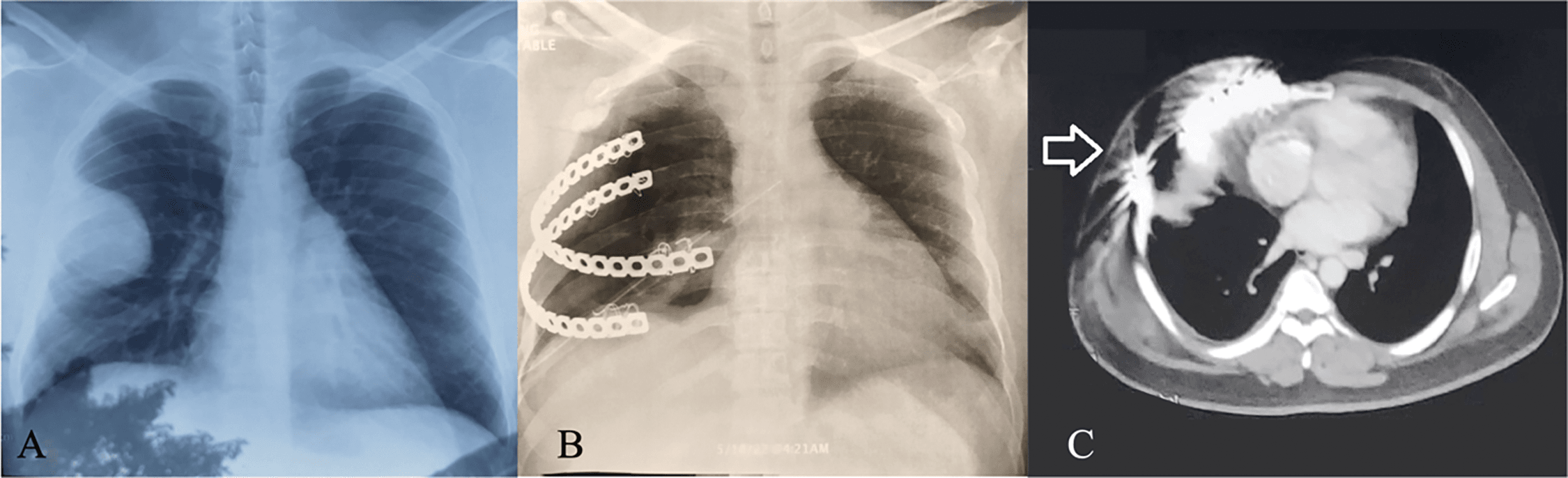
Figure 1. Radiograph posteroanterior view showing a mass on the right anterior chest wall. (A) Chest radiograph, Preoperative; (B) Chest radiograph, Postoperative; (C) Chest computed tomography, Postoperative.
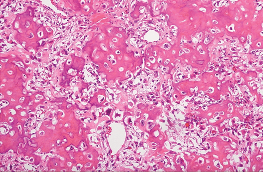
Figure 2. Pathological diagnosis based on H&E-stained sections. H&E, hemtoxylin and eosin.
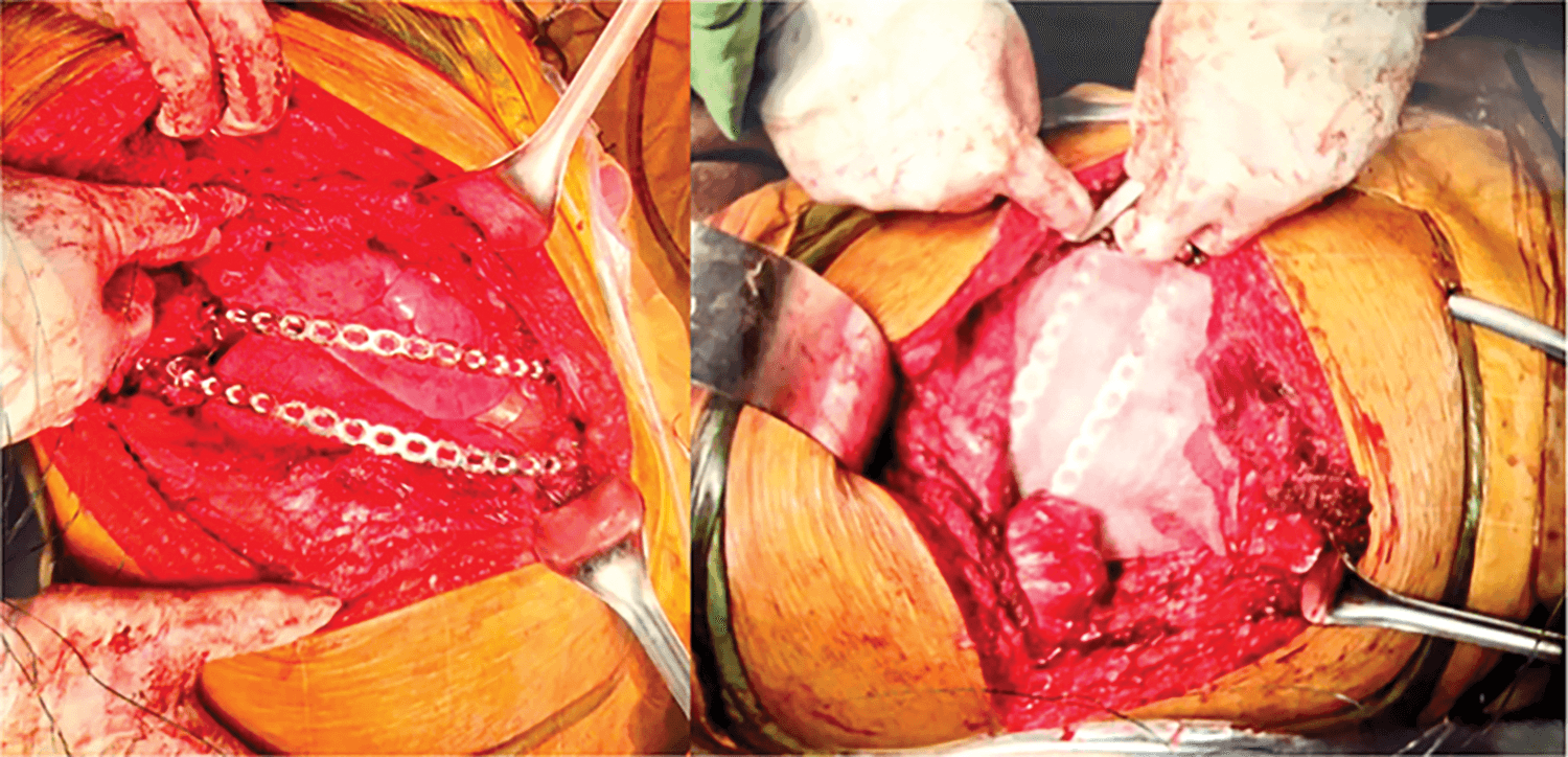
Figure 3. Intraoperative chest wall reconstruction.
At 1-month follow up, chest X-ray (Fig. 1B) and CT scan (Fig. 1C) showed interval resection of the fourth rib mass with metallic prostheses replacing the right fourth and fifth ribs. The mass at the vertebral end of the third rib remained unchanged. Imaging also showed decreased right lung volume, elevated right hemidiaphragm, and mild pleurodiaphragmatic adhesion. The patient received six cycles of adjuvant chemotherapy with cisplatin (100 mg/m2) and doxorubicin (75 mg/m2) every 21 days. Only grade 1 adverse effects were observed, and treatment was completed successfully. A follow-up chest CT scan 1 month after chemotherapy completion showed no recurrence, as well as stable placement of rib implants.
The patient remains asymptomatic and in good condition, expressing satisfaction with the successful tumor resection and completion of chemotherapy. Follow-up visits are scheduled every 3 months, with continued surveillance planned for a total duration of 5 years.
Discussion
This case illustrates an incidental finding of low-grade fibroblastic osteosarcoma in an asymptomatic adult male, which accounts for only 1.25% of all osteosarcoma cases.9 Diagnosis was confirmed through imaging and biopsy, followed by successful en bloc resection and adjuvant chemotherapy. The patient recovered fully, with no recurrence on follow-up. This highlights the importance of early detection and comprehensive management even in atypical presentations.
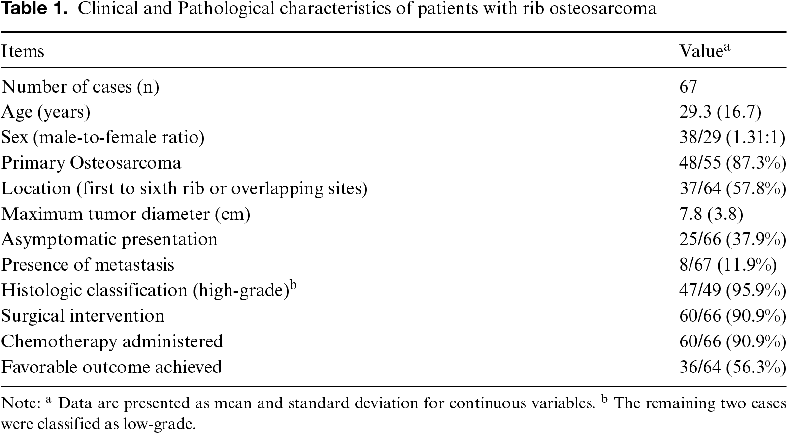
A PubMed search using the keywords “osteosarcoma” and “rib” yielded a summary of the clinical and pathological characteristics of 67 patients diagnosed with rib osteosarcoma, including the present case (Table 1).10–13 The mean age was 29.3 years (standard deviation: 16.7), with a male-to-female ratio of 1.31:1 (38 males, 29 females). Primary osteosarcoma accounted for 87.3% (48/55) of cases, with tumors most commonly located in the first to sixth ribs or overlapping rib regions (57.8%, 37/64). The average maximum tumor size was 7.8 cm (standard deviation: 3.8). Interestingly, 37.9% (25/66) of patients were asymptomatic at diagnosis. Metastatic disease was reported in 11.9% (8/67). High-grade histologic subtypes were identified in 95.9% (47/49) of cases. Surgical resection and chemotherapy were each performed in 90.9% (60/66) of patients, with favorable outcomes achieved in 56.3% (36/64).
Low-grade intraosseous osteosarcoma is a rare subtype of osteosarcoma that comprised 5.1% of cases in this literature review. It is characterized by osteoid or immature bone formation. For rib and other chest wall tumors, wide surgical excision is the primary treatment modality, which ensures clear margins and reduces recurrence. When large sections are removed, reconstruction is required to maintain respiratory function. Complete resection of the primary tumor and any metastases is critical for a potential cure, emphasizing the importance of preserving function.
Chemotherapy, often administered before and after the surgery, typically includes doxorubicin and cisplatin due to their effectiveness and tolerability, as recommended by the National Comprehensive Cancer Network.14 While the 5-year survival rates for rib osteosarcoma remain modest, strategies such as dose intensification are under investigation. Adverse effects may include myelosuppression, renal dysfunction, mucositis, and cardiotoxicity, although not all patients are affected.15 Long-term follow-up is essential, with studies like EURAMOS-1 showing favorable outcomes in nonmetastatic cases after complete resection.16 The poorer prognosis of this condition is linked to factors such as older age, male sex, large tumor size, axial location, poor chemotherapy response, or absence of surgery.17
This case report has several limitations. First, the follow-up duration is relatively short, which limits the evaluation of long-term oncologic outcomes, such as recurrence or survival. Second, functional outcomes, including postoperative pulmonary function and quality of life, were not formally assessed, which may reduce the clinical applicability of the findings. Additionally, advanced reconstruction techniques, such as 3D printing, were not available due to institutional limitations; therefore, a metallic rib prosthesis was selected based on its availability and suitability.
Conclusion
This case highlights a rare, asymptomatic presentation of low-grade osteosarcoma in the rib, discovered incidentally on routine imaging. Management with en bloc resection, reconstruction, and adjuvant chemotherapy led to a favorable outcome. Rib osteosarcoma remains uncommon and often lacks specific symptoms. This fact underscores the importance of early detection, complete surgical removal, and ongoing surveillance for optimal prognosis.
Acknowledgments
We appreciate the patient’s cooperation in data collection.
Funding Source
No financial support was sought from any source.
Author Contributions
G.B. was responsible for data curation, data interpretation, and drafting of the original manuscript. G.B. and M.A. made substantial contributions to revising the manuscript draft. All authors have read and approved the manuscript and agree with its content and data.
Data Availability
The datasets used in the current study can be obtained from the corresponding author upon reasonable request.
Ethical Statement
This article does not involve the participation of any animals. The patient provided written informed consent for the publication of this report and accompanying images.
Conflict of Interest
The authors report no conflicts of interest in this work.
Supplemental Information
Supplemental information for this article can be found online at https://sup.jclinque.com/api/articles/72/download-suppl.
References
[1] Kim C, Davis LE, Albert CM, Samuels B, Roberts JL, Wagner MJ. Osteosarcoma in pediatric and adult populations: are adults just big kids? Cancers (Basel). October 19, 2023;15(20):5044. doi:10.3390/cancers15205044.
[2] Cole S, Gianferante DM, Zhu B, Mirabello L. Osteosarcoma: a surveillance, epidemiology, and end results program-based analysis from 1975 to 2017. Cancer. June 1, 2022;1(11):2107–2118. doi:10.1002/cncr.34163.
[3] Cosci I, Del Fiore P, Mocellin S, Ferlin A. Gender differences in soft tissue and bone sarcoma: a narrative review. Cancers. 2024;16(1):201. doi:10.3390/cancers16010201.
[4] Bryan JN. Updates in osteosarcoma. Vet Clin North Am Small Anim Pract. May 2024;54(3):523–539. doi:10.1016/j.cvsm.2023.12.007.
[5] Dimayuga C. Frequency of primary malignant bone tumors in the Philippine General Hospital from 1985–1989. Acta Medica Philippina. 1992;2(6):71–74.
[6] Greenwood AC, Arora RD, Shaikh H. Osteosarcoma (Osteogenic Sarcoma). In: StatPearls. Treasure Island (FL): StatPearls Publishing; December 11, 2024.
[7] DePalma M, Gupta S, Nguyen J, Talwar D, Arkader A, Wells L. Do not miss the tumor: a novel presentation of osteosarcoma. Case Rep Pediatr. 2021;2021(4):5531238. doi:10.1155/2021/5531238.
[8] Menendez N, Epelman M, Shao L, Douglas D, Meyers AB. Pediatric osteosarcoma: pearls and pitfalls. Seminars in Ultrasound, CT and MRI. February 1, 2022;43(1):97–114. doi:10.1053/j.sult.2021.05.010.
[9] Wardoyo S, Kamal AF, Furqon MA, Grantomo J, Hutami WD. Osteosarcoma of the rib: a challenge of diagnosis and surgical intervention: a case report. Int J Surg Case Rep. April 2021;81(3):105777. doi:10.1016/j.ijscr.2021.105777.
[10] Hu X, Bao T, Yan C, Zhu Y, Zheng X. A case report of rib osteosarcoma and literature review. Clin Respir J. November 2023;17(11):1182–1189. doi:10.1111/crj.13686.
[11] Iwatsu J, Yoshida S, Watanuki M, et al. Clinical characteristics and prognosis of patients with incidentally discovered chest wall sarcoma compared with those of symptomatic patients. Jpn J Clin Oncol. August 14, 2024;54(8):911–916. doi:10.1093/jjco/hyae059.
[12] Bodmer N, Hecker-Nolting S, Friedel G, et al. Primary osteosarcoma of the ribs: a report from the cooperative osteosarcoma study group. Cancer. June 15, 2023;129(12):1895–1903. doi:10.1002/cncr.34744.
[13] Bae K, Moon JI, An HJ, Jung JJ, Jeon KN. High-grade surface osteosarcoma of the rib mimicking a neurogenic tumor: radiologic and pathologic findings. Diagnostics (basel). September 8 2023;13(18):2880. doi:10.3390/diagnostics13182880.
[14] NCCN Clinical Practice Guidelines in Oncology. Bone cancer. https://www.nccn.org/guidelines/guidelines-detail?category=1&id=1418. (Published February 2025, Accessed on March 15, 2025).
[15] Langer T, Stöhr W, Paulides M, et al. Prospective multicenter registration of major late sequelae in sarcoma patients using the Late Effects Surveillance System (LESS). Klin Padiatr. May–June 2005;217(3):176–181. doi:10.1055/s-2005-836503.
[16] Smeland S, Bielack SS, Whelan J, et al. Survival and prognosis with osteosarcoma: outcomes in more than 2000 patients in the EURAMOS-1 (European and American Osteosarcoma Study) cohort. Eur J Cancer. March 2019;109(1):36–50. doi:10.1016/j.ejca.2018.11.027.
[17] Xin S, Wei G. Prognostic factors in osteosarcoma: a study level meta-analysis and systematic review of current practice. J Bone Oncol. April 2020;21:100281. doi:10.1016/j.jbo.2020.100281.
 |
Copyright: © 2025 Batucan and Ando. This work is licensed under a Creative Commons Attribution 4.0 International License, which permits unrestricted use, distribution, and reproduction in any medium, provided the original work is properly cited. |