| Journal of Clinical Question. 2025; 2(1): e60 https://doi.org/10.69854/jcq.2025.0002 Advance access publication date 27 February 2025 |
 |
Case Report
Management with Conservative Gastrointestinal Decompression and Intravenous Immunoglobulin Relieved Intestinal Obstruction in Kawasaki Disease: A Case Report
Yuta Baba1, Yumeng Zhang 2,*, Yuri Ishida1, Akira Iwanaga1, Aiko Kumamoto1, Takeshi Yamanouchi3, Shinji Nishimura1
1Department of Pediatrics, Saga-Ken Medical Centre Koseikan, Saga, Japan.
2Department of Pediatrics, Faculty of Medicine, Saga University, Saga, Japan.
3Department of Pediatric Surgery, Faculty of Medicine, Saga University, Saga, Japan.
*Corresponding Author: e-mail: yumeng1990228@gmail.com
Submitted: January 19, 2025 Accepted: February 27, 2025
Clinical Question Box
What is the appropriate management for jejunal obstruction with bilious vomiting in a pediatric patient diagnosed with Kawasaki disease?
Incomplete jejunal obstruction in Kawasaki disease can be effectively managed through conservative measures, including intestinal decompression combined with intravenous immunoglobulin therapy and aspirin administration. It is essential to recognize the potential association between Kawasaki disease and ileus to avoid unnecessary surgical interventions, particularly in patients presenting with atypical symptoms.
Abstract
Background: Kawasaki disease (KD) is an acute febrile vasculitis that predominantly affects children under 5 years of age. Although gastrointestinal symptoms are common, severe complications, such as intestinal obstruction, are rare but clinically significant. Case Presentation: A 1-year and 7-month-old girl diagnosed with KD developed transient intestinal obstruction, presenting with bilious vomiting and hypochloremic alkalosis. Despite initial treatment with intravenous immunoglobulin (IVIG) and aspirin (ASA), her gastrointestinal symptoms worsened. Gastrointestinal imaging revealed an incomplete jejunal obstruction. Conservative management with intestinal decompression resolved the obstruction without surgical intervention, and the patient fully recovered without coronary complications. Conclusion: Intestinal obstruction in KD is rare but should be recognized, particularly in atypical presentations. Conservative approaches, including intestinal decompression combined with IVIG and ASA administration, can be effective alternatives to surgical interventions.
Keywords: Kawasaki disease, intestinal obstruction, intestinal decompression, intravenous immunoglobulin, acute abdominal symptoms, case report
Introduction
Kawasaki disease (KD) is an acute febrile systemic vasculitis that predominantly affects children under 5 years of age.1 It is most prevalent in Asian countries, with incidence rates ranging from 82.8 to 199.7 per 100,000 children under 5 years old, compared to lower rates of 10 to 20 per 100,000 in non-Asian regions, suggesting a potential genetic predisposition among Asian populations.2,3 KD is characterized by fever accompanied by specific clinical features, such as bilateral conjunctival hyperemia, polymorphous exanthema, oral mucosal changes, peripheral extremity changes, and cervical lymphadenopathy.4 The disease primarily affects medium-sized arteries, making early diagnosis and treatment essential to prevent coronary artery complications, which are the most critical aspects of the disease.5
Gastrointestinal symptoms, such as diarrhea, vomiting, and abdominal pain, are frequently observed in KD.6,7 Most symptoms are mild and transient.7 However, severe gastrointestinal complications, such as intestinal obstruction and ischemic colitis, are rare but can occur in KD, posing significant diagnostic and therapeutic challenges.8 Here, we present a unique case of a 1-year and 7-month-old girl diagnosed with KD who developed transient jejunal obstruction with bilious vomiting during her illness.
Case Presentation
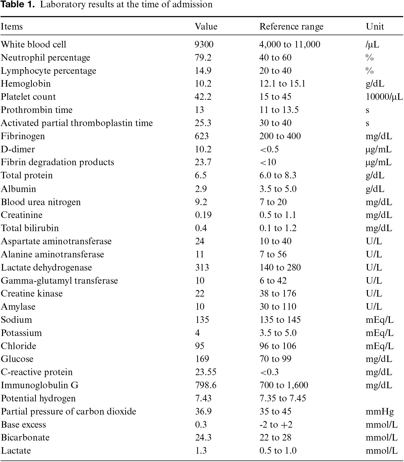
A 1-year-and-7-month-old girl with a fever ranging from 38°C to 40°C was initially diagnosed with pharyngitis and treated with faropenem from day 1 to 3. On day 6, she developed reddened lips, conjunctival hyperemia, and polymorphous exanthema. The antibiotic treatment was then switched to clarithromycin, but there was still no response. On day 7, she began experiencing nausea. On the eighth day of persistent fever, she was referred to our hospital. She had a bowel movement every 2 days. She is not a picky eater and had no contact with well water or pets. Upon arrival at our hospital, her vital signs were stable: body temperature 37.6°C, heart rate 148/min, blood pressure 124/76 mmHg, RR 28/min, and SpO2 97%. Physical examination revealed bilateral conjunctival hyperemia, reddened lips, polymorphous exanthema, and indurative edema. There was no abdominal distension or tenderness, and bowel sounds were normal. No hepatomegaly was detected. Laboratory examinations showed a white blood cell count of 9.3 × 10⁹/L, with neutrophil predominance (79.2%), a hemoglobin level of 102 g/L, and a platelet count of 422 × 109/L. Liver enzyme and bilirubin levels were within normal ranges. C-reactive protein was elevated to 23.55 mg/dL (Table 1). Rapid antigen tests for streptococcus and adenovirus were negative, and blood and stool cultures did not identify any pathogenic bacteria. Echocardiography revealed no abnormalities in the coronary arteries.
The patient was diagnosed with KD, and treatment was initiated with aspirin (ASA) at a dose of 30 mg/kg per day. Intravenous immunoglobulin (IVIG, 2 g/kg) was started on the ninth day of KD. As shown in Fig. 1, the typical symptoms of KD, such as reddened lips and polymorphous exanthema, improved after treatment. C-reactive protein gradually decreased following IVIG, and membranous desquamation was observed on day 12. However, after admission, the patient’s vomiting worsened, progressing to bilious vomiting, accompanied by hypochloremic alkalosis (blood gas test: pH 7.622, HCO3− 40.2 mmol/L, Cl- 86 mmol/L). Fasting management failed to alleviate the bilious vomiting. An abdominal X-ray revealed decreased bowel gas shadowing, and an abdominal ultrasound showed proximal small intestine dilation, edema of the intestinal wall, and scattered lymph nodes under 10 mm in size throughout the abdominal cavity (Figs. 2A–2C). On the 11th day of KD, abdominal computed tomography revealed proximal jejunum dilation and fluid retention. The dilated jejunum exhibited obstruction at the indicated site, while the intestine distal to this segment appeared to collapse (Fig. 2D). An elemental diet (ED) tube was inserted on the same day, and gastrointestinal imaging was performed. Contrast medium injection showed no flow beyond the dilated intestinal segment. Even with repositioning, the contrast medium did not pass through. However, 3 hours later, follow-up X-rays revealed that the contrast medium had reached the distal intestine, suggesting that the obstruction was incomplete (Fig. 3).
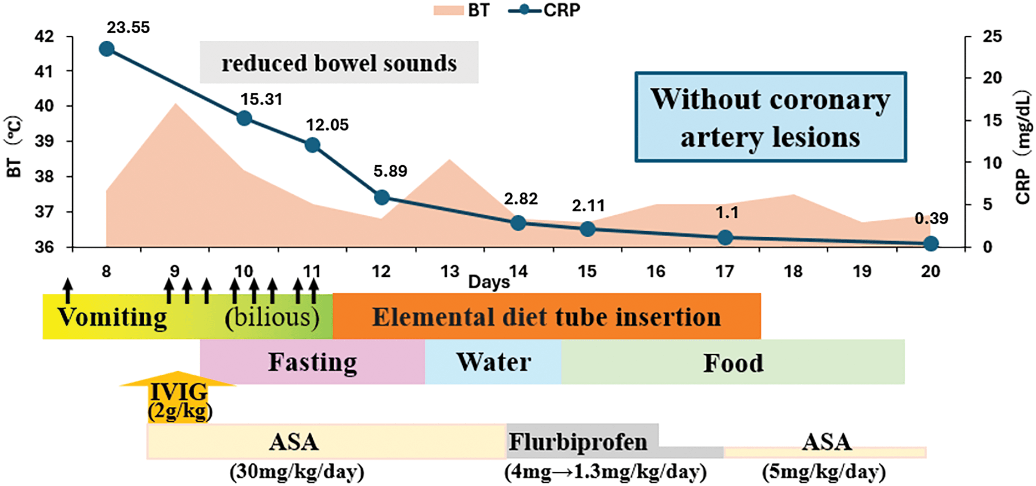
Figure 1. Clinical course of the patient. BT: body temperature; CRP: C-reactive protein; IVIG: intravenous immunoglobulin; ASA: aspirin.
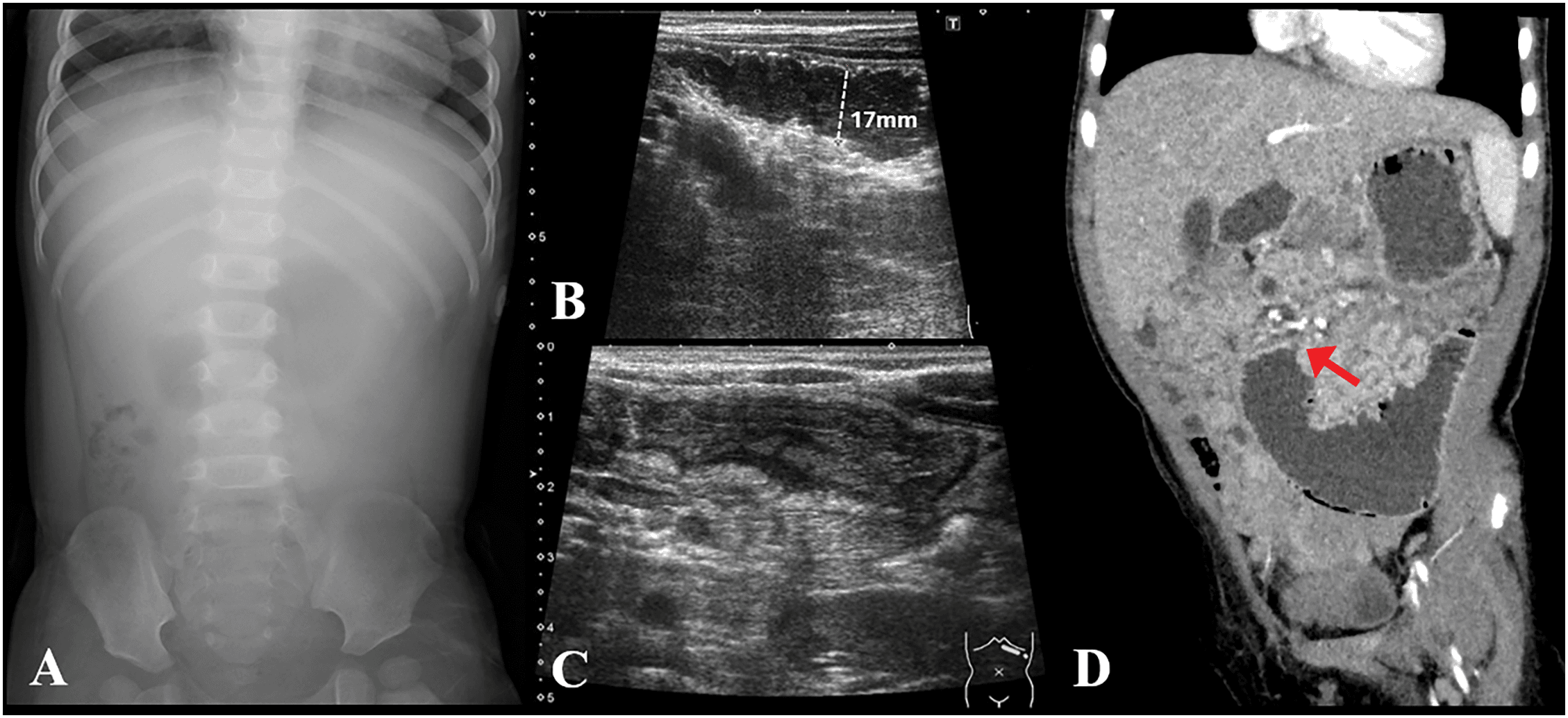
Figure 2. Imaging findings of the patient. (A) Abdominal radiograph taken on day 9. (B, C) Abdominal ultrasound performed on day 10. (D) Computed tomography scan of the abdomen on day 11.
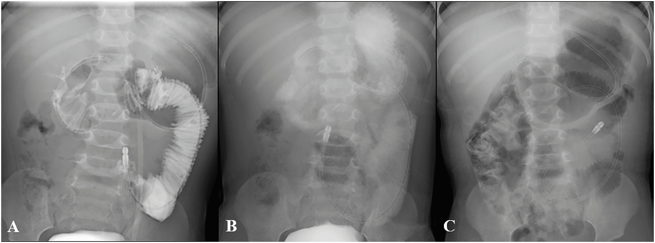
Figure 3. Contrast study findings following elemental diet tube placement. (A) Abdominal radiograph confirming the successful placement of the elemental diet tube. The contrast study showed no flow of contrast medium beyond the dilated intestinal segment. (B) Follow-up X-ray revealed that the contrast medium had reached the distal intestine 3 hours later. (C) Follow-up X-ray the day after the administration of the elemental diet tube.
Gastrointestinal decompression was performed using the ED tube, and the patient’s condition was monitored with continuous drainage. The drainage volume gradually decreased, and the symptoms of KD improved. Two days later, oral intake was resumed as the intestinal gas pattern improved and vomiting subsided. After an additional 2 days, regular eating was restarted without further vomiting. With the intestinal obstruction resolved, the ED tube was removed. As the KD symptoms continued to improve, the antiplatelet agent dose was gradually reduced. Subsequent clinical progress was favorable, without recurrence of vomiting and evidence of coronary artery dilatation. The patient and her parents were satisfied with the treatment, and the patient was discharged in good condition.
Discussion
This case underscores an unusual gastrointestinal complication of KD: transient jejunal obstruction. The incidence of acute abdominal symptoms in KD is reported to be approximately 4.4%. Among patients with these symptoms, the prevalence of intestinal obstruction increases significantly, reaching as high as 25.6%.9,10 Due to the typical symptoms, this case was promptly diagnosed and effectively treated with IVIG and ASA. The patient’s bilious vomiting and hypochloremic alkalosis required close monitoring and timely intervention. ED tube drainage provided symptomatic relief and facilitated the resolution of the obstruction, successfully preventing the need for invasive surgery, with no coronary artery complications detected.
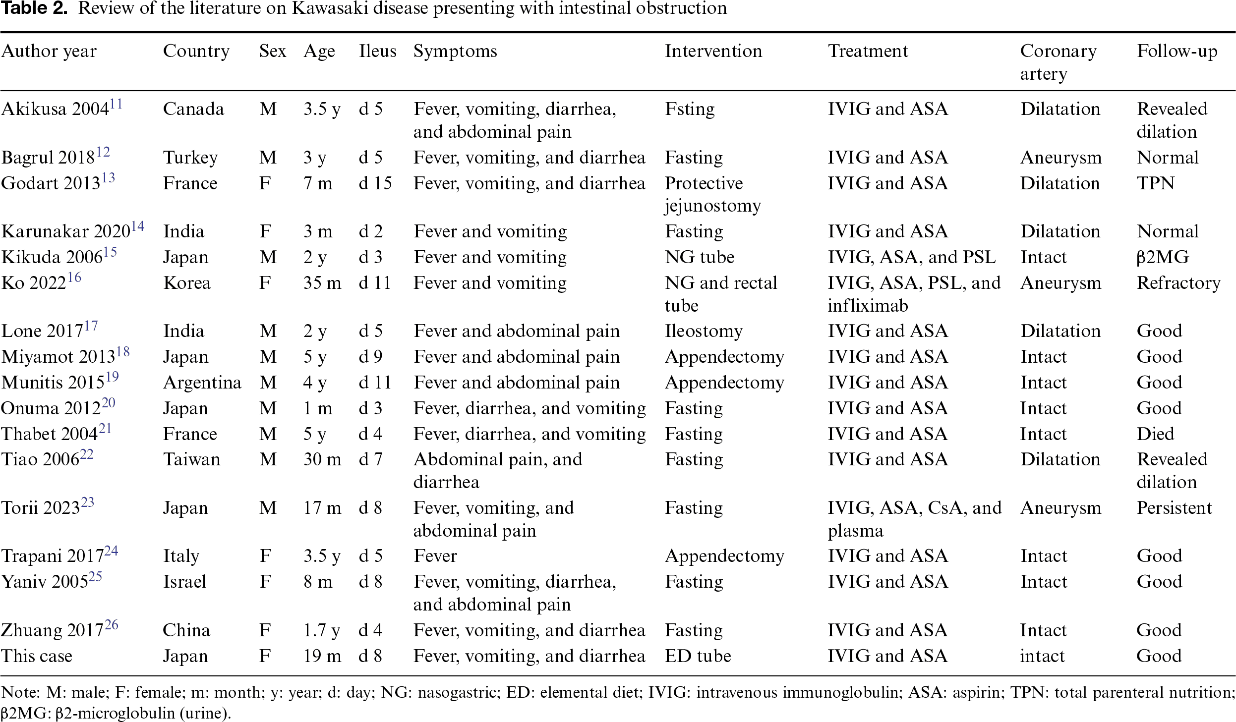
A literature review of studies on KD patients with intestinal obstruction over the past 20 years was conducted and summarized in Table 2. Seventeen cases, including our case, reported the presentation of intestinal obstruction in KD.11–26 The male-to-female ratio was 1.43, with a median age and interquartile range of 19 months (7, 35) in females and 33 months (22.2, 51) in males. The onset of intestinal obstruction occurred at a median of day 8 (4, 11) in females and day 5 (3.75, 8.25) in males. Fever, vomiting, diarrhea, and abdominal pain were the most frequently observed symptoms. In particular, bilious vomiting and diffuse abdominal tenderness are significant indicators. Additionally, a follow-up X-ray is essential, as it can promptly detect dilated bowel loops and intestinal wall thickening. All cases were treated with IVIG and ASA. Gastrointestinal symptoms improved in nine cases with fasting alone. Two cases, similar to ours, were managed with a nasogastric tube. Five cases (29.4%) underwent surgery before the definitive diagnosis of KD, with 60% of these surgeries being appendectomies. Abnormalities in the coronary arteries were observed in eight patients (47.1%), a notably higher incidence than typically seen in KD. Of these, half resolved to normal, two showed coronary artery dilation, and the remaining two developed aneurysms, which exhibited no changes during follow-up.
Diagnosing patients with KD presenting with acute abdominal symptoms is challenging, particularly in atypical KD cases with persistent abdominal symptoms only. The exact mechanism of intestinal obstruction in KD remains unclear. The jejunum has a rich vascular supply and a relatively high metabolic rate, making it more susceptible to ischemic or inflammatory insults. It is postulated that vasculitis-related inflammation affecting mesenteric arteries could impair bowel motility or lead to localized edema and intestinal wall thickening, contributing to obstruction.27 Recognizing and considering the possibility of KD complicated with intestinal obstruction is crucial for surgeons and pediatricians. Clinicians should remain vigilant for abdominal symptoms and consider KD-related complications to ensure timely diagnosis and appropriate management. One study even reported appendicitis as the presenting manifestation of KD,28 further complicating the situation. A conservative approach using an ED tube may be useful in relieving symptoms in patients with suspected KD while awaiting the development of other typical symptoms of KD.
Conclusion
This case highlights a rare but clinically significant gastrointestinal complication of KD, specifically transient jejunal obstruction. Early recognition and conservative management with gastrointestinal decompression using an ED tube for drainage, combined with standard treatments including IVIG and ASA, were crucial in achieving a successful outcome without requiring invasive surgery or leading to coronary complications. Nevertheless, clinicians should remain vigilant when assessing atypical presentations of KD, especially in patients with persistent gastrointestinal symptoms only, to ensure efficient and effective intervention.
Acknowledgment
We would like to extend our gratitude to the patient for cooperation in data collection.
Funding Source
No financial support was provided.
Author Contributions
Y.B. and Y.Z. drafted the original manuscript. Y.B. and Y.Z. were responsible for data curation and interpretation. Y.I., A.I., A.K., T.Y., and S.N. substantially contributed to the manuscript draft. All authors have read and approved the manuscript and agree with the content and data.
Data Availability Statement
The datasets used in the current study are available from the corresponding author upon reasonable request.
Ethical Statement
The article does not involve the participation of any animals. The patient gave written informed consent to the publication of this report and the accompanying images.
Conflict of Interest
The authors report no conflicts of interest in this work.
References
[1] Bhatta M, Baniya A, Koirala D, Bhattarai K, Shah A. Dilemma in diagnosing incomplete Kawasaki disease in a resource limited setting. Ann Med Surg (Lond). 2023;85(11):5809–5812. doi:10.1097/MS9.0000000000001380.
[2] Rowley AH, Shulman ST. The epidemiology and pathogenesis of Kawasaki disease. Front Pediat. 2018;6:374. doi:10.3389/fped.2018.00374.
[3] Son MBF, Newburger JW. Kawasaki disease. Pediatr Rev. 2018;39(2):78–90. doi:10.1542/pir.2016-0182.
[4] Kuo HC. Diagnosis, progress, and treatment update of Kawasaki disease. Int J Mol Sci. 2023;24(18):13948. doi:10.3390/ijms241813948.
[5] Broderick C, Kobayashi S, Suto M, Ito S, Kobayashi T. Intravenous immunoglobulin for the treatment of Kawasaki disease. Cochrane Database Syst Rev. 2023;1(1):Cd014884. doi:10.1002/14651858.CD014884.pub2.
[6] Yang P, Mao Z, Sun M, Guo J. Clinical features analysis of Kawasaki disease with abdominal symptoms as the first manifestation. Eur J Pediatr. 2023;182(9):4049–4057. doi:10.1007/s00431-023-05086-x.
[7] McCrindle BW, Rowley AH, Newburger JW, et al. Diagnosis, treatment, and long-term management of Kawasaki disease: a scientific statement for health professionals from the American Heart Association. Circulation. 2017;135(17):e927–e999. doi:10.1161/CIR.0000000000000484.
[8] Fabi M, Corinaldesi E, Pierantoni L, et al. Gastrointestinal presentation of Kawasaki disease: a red flag for severe disease? PLoS One. 2018;13(9):e0202658. doi:10.1371/journal.pone.0202658.
[9] Miyake T, Kawamori J, Yoshida T, Nakano H, Kohno S, Ohba S. Small bowel pseudo-obstruction in Kawasaki disease. Pediatr Radiol. 1987;17(5):383–386. doi:10.1007/BF02396613.
[10] She X, Chen J, Zhou YN, Guo J, Zhao FH, Yi C. Kawasaki disease associated acute abdomen: most require no surgery. J Inflamm Res. 2023;16:5157–5162. doi:10.2147/JIR.S434982.
[11] Akikusa JD, Laxer RM, Friedman JN. Intestinal pseudoobstruction in Kawasaki disease. Pediatrics. May 2004;113(5):e504–e506. doi:10.1542/peds.113.5.e504.
[12] Bagrul D, Karadeniz EG, Koca S. Gastrointestinal involvement in Kawasaki disease: a case report. Cardiol Young. August 2018;28(8):1070–1073. doi:10.1017/S1047951118000847.
[13] Godart F, Bakhti OM, Bonnevalle M. Intestinal ischaemia as a severe presentation of Kawasaki disease leading to short-bowel syndrome. Cardiol Young. June 2014;24(3):567–570. doi:10.1017/S1047951113000966.
[14] Karunakar P, Gunasekaran D, Ramamoorthy JG, et al. Kawasaki disease presenting with intestinal pseudo-obstruction. Indian J Pediatr. July 2020;87(7):569–570. doi:10.1007/s12098-020-03298-9.
[15] Kikuda Y. A case of Kawasaki disease complicated by paralytic ileus and hemophagocytic syndrome. Jpn J Pediatr. 2006;59(3):463–470. http://id.ndl.go.jp/bib/7847690.
[16] Ko H, Byun JH, Kim G, Lee HD, Kim TH. Kawasaki disease with intestinal pseudo obstruction and neurogenic bladder. Pediatr Int. January 2022;64(1):e15038. doi:10.1111/ped.15038.
[17] Lone YA, Menon J, Menon P, et al. Kawasaki disease presenting as acute intestinal obstruction. J Indian Assoc Pediatr Surg. July–September 2017;22(3):170–172. doi:10.4103/0971-9261.207632.
[18] Miyamoto K, Yamazaki Y, Okamoto K, Tsuboi T, Hirao J, Arisaka O. Kawasaki disease: relationship between acute surgical abdomen and cytokine profiles. Pediatr Infect Dis. November 2013;32(11):1299. doi:10.1097/INF.0b013e31829ece3d.
[19] García Munitis P, Ves Losada J, Montali C. Acute abdomen at onset of incomplete and atypical Kawasaki disease: case report. Arch Argent Pediatr. 2015;113(2):e88–93. doi:10.5546/aap.2015.e88.
[20] Oonuma N, Oki A, Sekijima T, Kagimoto S, Ichihashi K. A case of Kawasaki disease complicated by paralytic ileus in a 1-month-old infant. Jpn J Pediatr. September 2012;65(9):2055–2060.
[21] Thabet F, Bellara I, Tabarki B, et al. Colite ischémique et syndrome d’hémophagocytose compliquant une maladie de Kawasaki. Archives de Pédiatrie. March 01, 2004;11(3):226–228. doi:10.1016/j.arcped.2003.12.017.
[22] Tiao MM, Huang LT, Liang CD, Ko SF. Atypical Kawasaki disease presenting as intestinal pseudo-obstruction. J Formos Med Assoc. March 2006;105(3):252–255. doi:10.1016/S0929-6646(09)60315-6.
[23] Torii A, Morishita G, Ogino K. A case of Kawasaki disease refractory to immunoglobulin therapy complicated by paralytic ileus requiring plasma exchange. Ann Kurashiki Central Hospita. 2023;85:115–119.
[24] Trapani S, Montemaggi A, Simonini G, Calabri GB, Messineo A, Resti M. Surgical abdomen with intestinal pseudo-obstruction as presenting feature of atypical Kawasaki disease. J Paediatr Child Health. November 2016;52(11):1032–1034. doi:10.1111/jpc.13284.
[25] Yaniv L, Jaffe M, Shaoul R. The surgical manifestations of the intestinal tract in Kawasaki disease. J Pediatr Surg. 2005;40(9):e1–e4. doi:10.1016/j.jpedsurg.2005.05.063.
[26] Zhuang Y, Zhou Y, Gu Y. Intestinal pseudo-obstruction as the presenting manifestation of Kawasaki disease. Indian J Pediatr. August 2017;84(8):647–648. doi:10.1007/s12098-017-2345-x.
[27] Kapoor S. Kawasaki’s disease: an often overlooked cause of intestinal pseudo-obstruction in children. Virchows Archiv. November 01, 2015;467(5):619–620. doi:10.1007/s00428-015-1844-2.
[28] Garnett GM, Kimball S, Melish ME, et al. Appendicitis as the presenting manifestation of Kawasaki disease. Pediatr Surg Int. May 2014;30(5):549–552. doi:10.1007/s00383-013-3439-9.
 |
Copyright: © 2025 Baba et al. This work is licensed under a Creative Commons Attribution 4.0 International License, which permits unrestricted use, distribution, and reproduction in any medium, provided the original work is properly cited. |