| Journal of Clinical Question, 2024, Vol. 1, No. 3, 159–166 https://doi.org/10.69854/jcq.2024.0021 Advance access publication date 31 December 2024 |
 |
Case Report
Long-term Follow-up of a Patient with Localized Bronchiectasis Complicated by Bronchial Aspergillosis After Bronchial Thermoplasty: A Case Report and Literature Review
Fumihiro Kashizaki 1,* and Yasutomo Sekido2
1Department of Respiratory Medicine, Yokohama Minami Kyosai Hospital, Yokohama, Japan.
2Department of Pathology, Isehara Kyodo Hospital, Tanaka, Isehara, Japan.
*Corresponding Author: e-mail: f.kashizaki@gmail.com
Submitted: November 17, 2024; Accepted: December 30, 2024
Clinical Question Box
What is the clinical course of a patient with postoperative bronchial aspergillosis after bronchial thermoplasty?
Allergic bronchopulmonary aspergillosis, chronic pulmonary aspergillosis, or an overlap of these conditions may arise, necessitating antifungal treatment, systemic corticosteroids, or both. Following these therapies, some patients may develop localized bronchiectasis, which can lead to chronic airway infections requiring further treatment strategies.
Abstract
Background: Bronchiectasis remains incompletely understood in terms of its causes, progression, and treatment strategies. It frequently arises as a secondary condition, underscoring the need to include it as a differential diagnosis during clinical evaluation. Case Presentation A 35-year-old woman with type 2 severe bronchial asthma presented with persistent symptoms two weeks after bronchial thermoplasty (BT). Chest computed tomography revealed bronchial wall thickening and bronchiectasis in the left B8 bronchus. Bronchoscopy showed cultures positive for Aspergillus fumigatus from bronchial lavage fluid, and transbronchial biopsy revealed hyphae. The patient was diagnosed with bronchial aspergillosis localized to the left B8 bronchus as a postoperative complication of BT. Treatment with voriconazole for six months led to gradual improvement in bronchial aspergillosis, with imaging showing resolution of bronchial wall thickening and a trend toward improvement in bronchiectasis. Persistent yellow sputum prompted repeat bronchoscopy, revealing negative Aspergillus fumigatus cultures but positive Staphylococcus aureus, indicating localized bronchiectasis with chronic airway infection. Symptoms partially improved with low-dose erythromycin after limited response to repeated amoxicillin/clavulanic acid. Asthma control also improved post-BT. Conclusions: Bronchial aspergillosis is a rare complication of BT that can progress to localized bronchiectasis and chronic airway infections, necessitating careful long-term follow-up. The disease course appears similar to other types of bronchiectasis, even when it develops as a complication of BT.
Keywords: Bronchiectasis, bronchial thermoplasty, bronchial aspergillosis, bronchial asthma, Aspergillus fumigatus, case report.
Introduction
Despite advancements in the treatment of severe bronchial asthma (BA)1,2 and allergic bronchopulmonary aspergillosis (ABPA),3 some patients have reported secondary bronchiectasis.4 The coexistence of bronchiectasis with these conditions poses significant challenges for disease management.5 While the underlying mechanisms driving bronchiectasis progression and its treatment strategies are not fully understood, various clinical trials have proposed the vicious vortex model and introduced novel therapeutic approaches.6–9 In this report, we present a rare case of bronchial aspergillosis following bronchial thermoplasty (BT), complicated by localized bronchiectasis and chronic airway infection. We review the existing literature on BT-related bronchial aspergillosis, including this case, and discuss potential treatment strategies for secondary localized bronchiectasis.
Case Presentation
A 35-year-old woman with a history of severe type 2 bronchial asthma (BA) was admitted to the hospital for persistent cough and purulent sputum, which worsened following the completion of BT. She had no significant medical history besides BA, including allergic bronchopulmonary mycosis, and was a non-smoker. Her home environment was observed to be humid, with occasional mold odors.
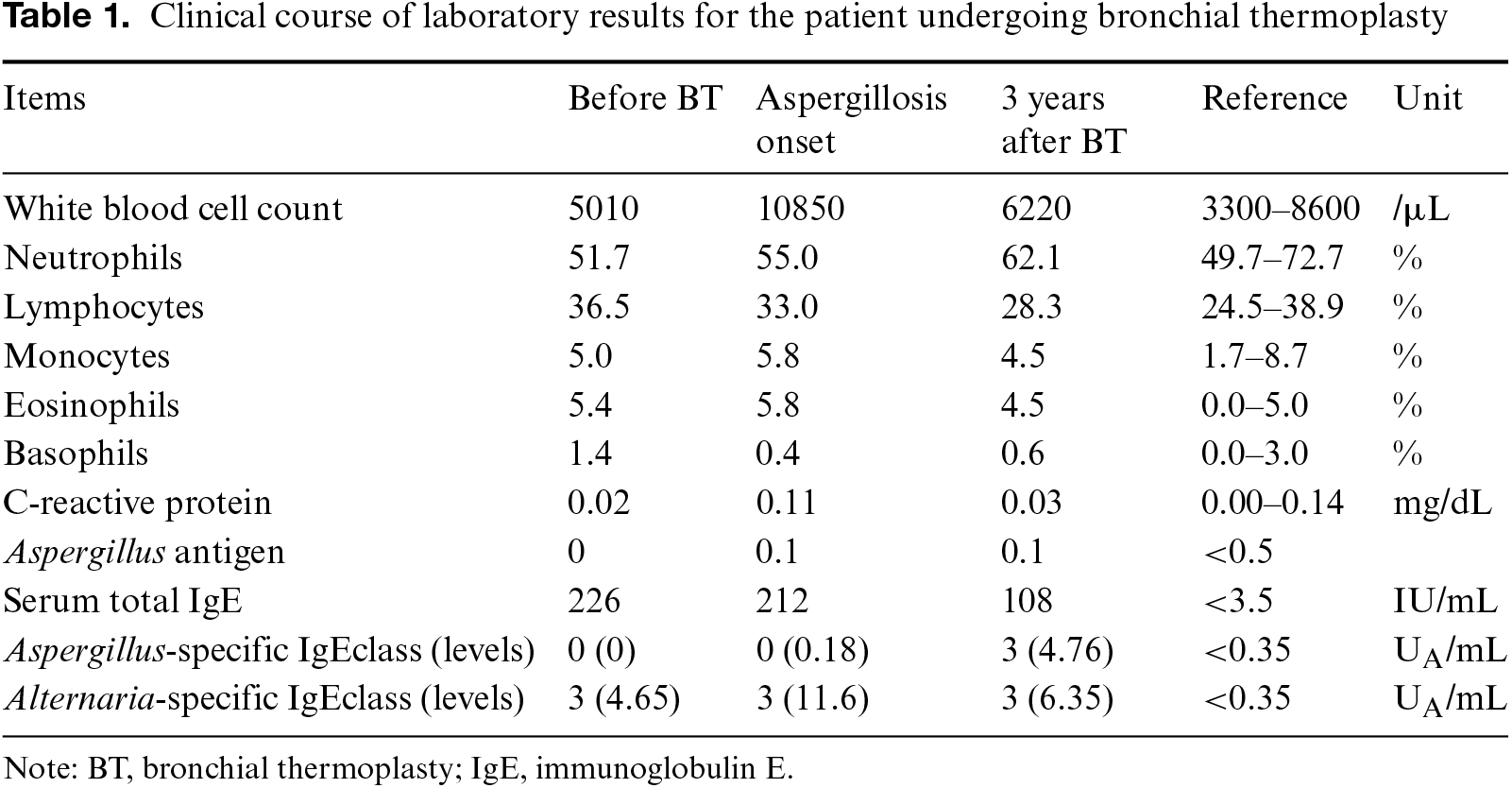
The patient’s asthma, classified as Global Initiative for Asthma step 5, was treated with high-dose inhaled corticosteroids, long-acting β2 agonists, long-acting muscarinic antagonists, and leukotriene receptor antagonists. Despite this regimen, she occasionally required oral corticosteroids for exacerbations, occurring once or twice per year. Three weeks before admission, she had completed three BT procedures, each accompanied by systemic steroid administration (prednisolone, 50 mg daily for 5 days) (Fig. 1). The first BT procedure was performed on the right lower lobe, the second on the left lower lobe, and the third on the bilateral upper lobes. During the third BT treatment, the left lower lobe was observed to reveal only airway mucosal changes that could be attributed to activation during the second BT treatment. Serum-specific immunoglobulin E (IgE) testing before BT revealed sensitization to Alternaria but was negative for Aspergillus (Table 1, Table S1). Physical examination at admission showed no abnormalities. Blood tests indicated an elevated white blood cell count and a slight increase in CRP compared to pre-BT levels.
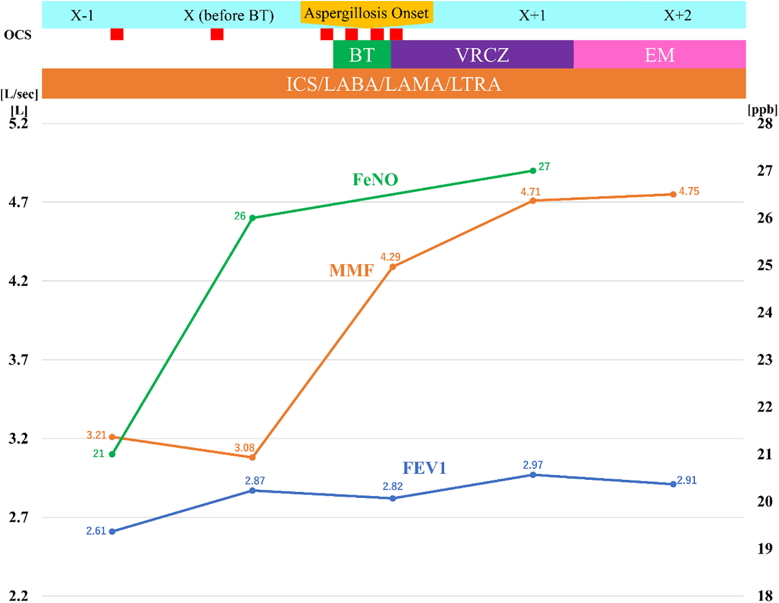
Figure 1: Clinical course and pulmonary function test findings. OCS; oral corticosteroid; BT, bronchial thermoplasty; EM, erythromycin; ICS, inhaled corticosteroids; LABA, long-acting β2 agonists; LAMA, long-acting muscarinic antagonists; LTRA, leukotriene receptor antagonists; FeNO, fractional exhaled nitric oxide; MMF, maximum mid-expiratory flow rate; FEV1, forced expiratory volume in one second.
Chest computed tomography (CT) revealed focal thickening of the left lower bronchial wall, bronchiectasis, consolidation, opacities, and changes consistent with prior BT (Fig. 2A). On suspicion of BT-related complication, a bronchoscopy was performed and revealed necrotic tissue and purulent mucus within the lumen, predominantly in B8 of the left lung along the treated areas in the lower lobe (Fig. 2D). Transbronchial biopsy reveals necrotic material containing fungal hyphae, which stain black with Grocott’s stain and display sharply branching structures (Figs. 3A–3C). Bronchial lavage fluid cultures confirmed the presence of Aspergillus fumigatus, prompting a diagnosis of bronchial aspergillosis. Treatment with voriconazole was initiated.
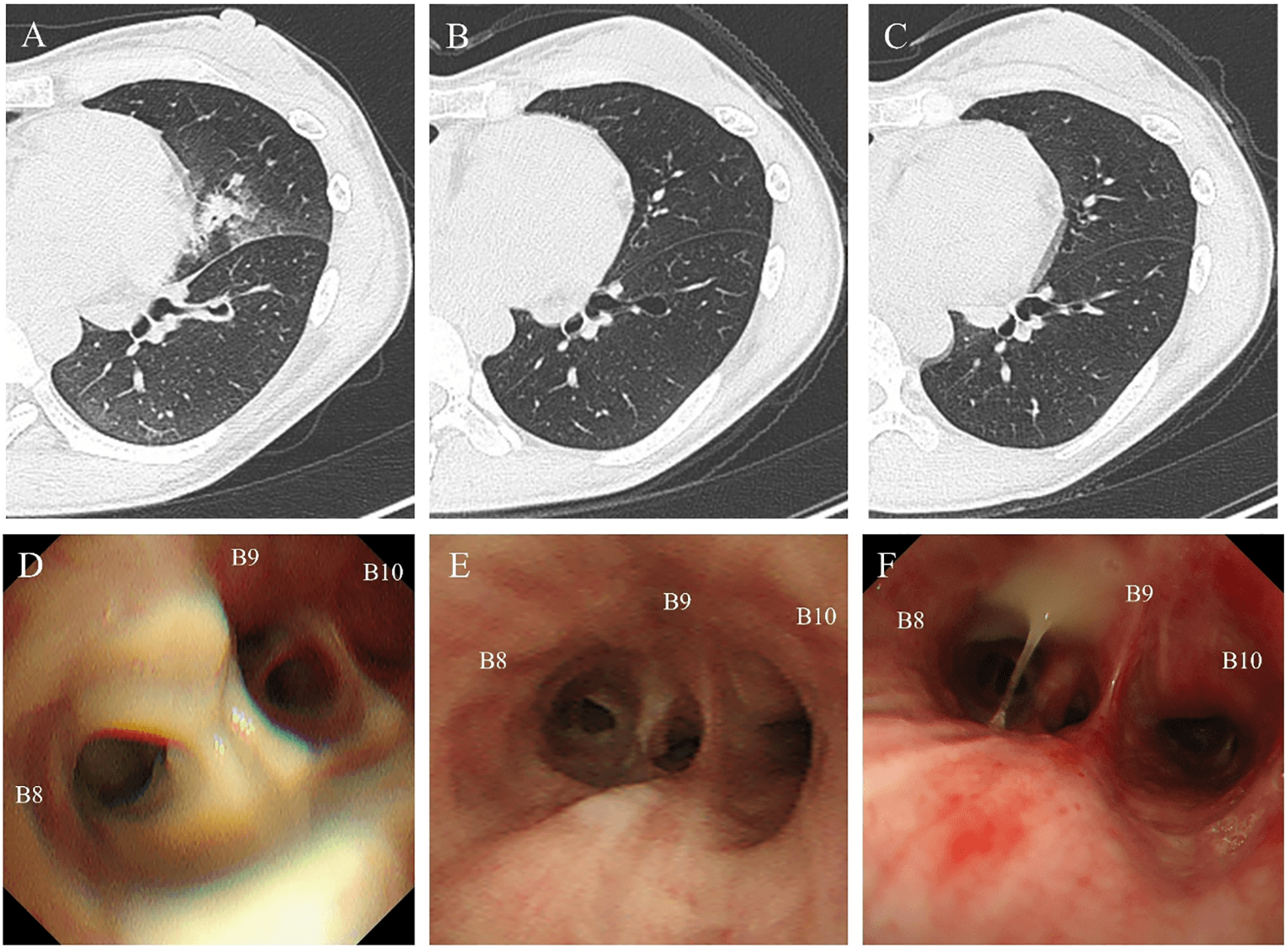
Figure 2: Patient’s computed tomography and bronchoscopy images. (A) Diagnosis: CT shows bronchial wall thickening, bronchiectasis, consolidation, opacities, and upper lobe changes; (B) Six months: CT shows improvement in all parameters except bronchiectasis; (C) Three years: CT shows near-complete resolution; (D) Diagnosis: Bronchoscopy shows necrotic tissue and purulent mucus in the left B8 bronchus; (E) Six months: Bronchoscopy shows resolved necrosis and bronchial scarring; (F) Three years: Bronchoscopy shows improved scarring; purulent sputum persists.
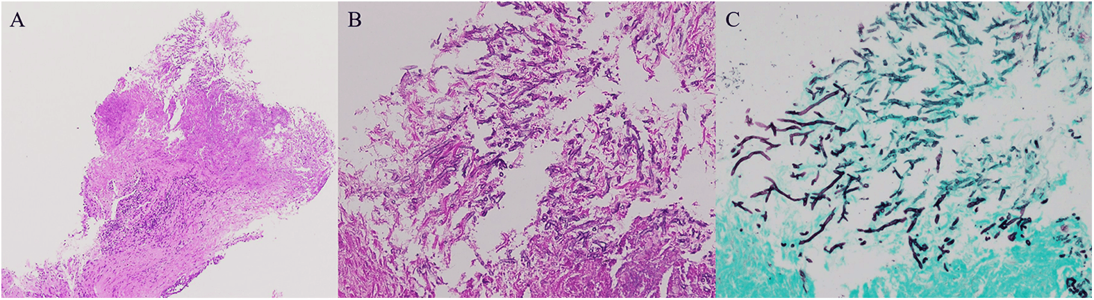
Figure 3: Patient’s pathological findings. Hematoxylin and eosin staining revealed necrotic material with inflammatory cell infiltration (A, ×40; B, ×200). The Grocott-stained specimen (C, ×200) demonstrates fungal hyphae with consistent width and black.
After six months of voriconazole treatment, intermittent purulent sputum persisted. Chest CT demonstrated improvement in bronchial wall thickening, consolidation, and opacities; however, bronchiectasis remained (Fig. 2B). Bronchoscopy showed bronchial wall scarring and the absence of necrotic tissue (Fig. 2E). Sputum cultures were negative for Aspergillus fumigatus but revealed Staphylococcus aureus. The patient was treated with amoxicillin/clavulanic acid several times due to infectious exacerbation of bronchiectasis. Consequently, the patient was switched to low-dose erythromycin (400 mg daily). After starting erythromycin, the purulent sputum significantly improved but still intermittently persisted. Chest CT revealed further improvement in bronchial wall thickening and bronchiectasis (Fig. 2C). Bronchoscopy showed ongoing improvement in bronchial wall scarring, although purulent sputum was still present (Fig. 2F). Sputum culture remained positive for Staphylococcus aureus. The patient’s asthma remained stable, including her pulmonary function (Fig. 1), but she required ongoing treatment for localized bronchiectasis.
Discussion
The patient developed aspergillosis as a postoperative complication following BT, which improved with voriconazole; however, localized bronchiectasis persisted. Subsequently, the patient experienced chronic lower respiratory tract infection caused by staphylococci. Bronchial aspergillosis has been reported as a postoperative complication in approximately 5% of BT cases.10,11 Including the present case, five patients with bronchial aspergillosis have been reported, none of whom were suspected to have ABPA before treatment.10,12–15 Separately, one additional case involved a patient in whom ABPA was suspected before treatment.16 We analyzed five cases (Table 2),10,12–15 excluding one where ABPA was suspected before BT.16 All patients were female. Each patient received 30–50 mg of prednisolone daily for approximately five days and developed bronchial aspergillosis localized to the lower lobe bronchus within six weeks of undergoing BT. Among the five cases, this case had the longest time to disease onset. However, it is possible that the bronchial aspergillosis was mild at the time of the third procedure BT, being regarded as post-BT airway mucosal changes during observation. Most cases are likely to develop within four weeks. In three of these cases,10,12,13,15 elevated Aspergillus-specific IgE levels were observed after the onset of aspergillosis, although none reported the presence of a clear mucus plug. In our two cases where Aspergillus antigen levels were measured before and after BT, the previously reported case10 showed a significant increase, while in the present case, the levels remained within the normal range. This pattern may suggest a combination of localized Aspergillus infection and ABPA-like conditions,17 potentially influenced by individual allergic predispositions and airway-destructive factors.18–20 While da Costa et al. described this condition as Aspergillus tracheobronchitis,13 we refer to it as bronchial aspergillosis to account for individual immune conditions and its diverse clinical presentations. Factors contributing to the development of bronchial aspergillosis following BT possibly include the short-term use of moderate to high doses of systemic corticosteroids, mucosal damage to the central airways (an environment favorable to Aspergillus growth), the lower lobe’s susceptibility to mucociliary impairment, and the patient’s home environment.12 Antifungal treatment was administered in all five reported cases, with one treated with itraconazole,14 three with voriconazole,10,12,15 and one transitioning from voriconazole to amphotericin B inhalation.13 In all cases, aspergillosis improved within one year, possibly due to the infection being localized. However, the long-term outcomes of bronchial aspergillosis after BT remain unknown.
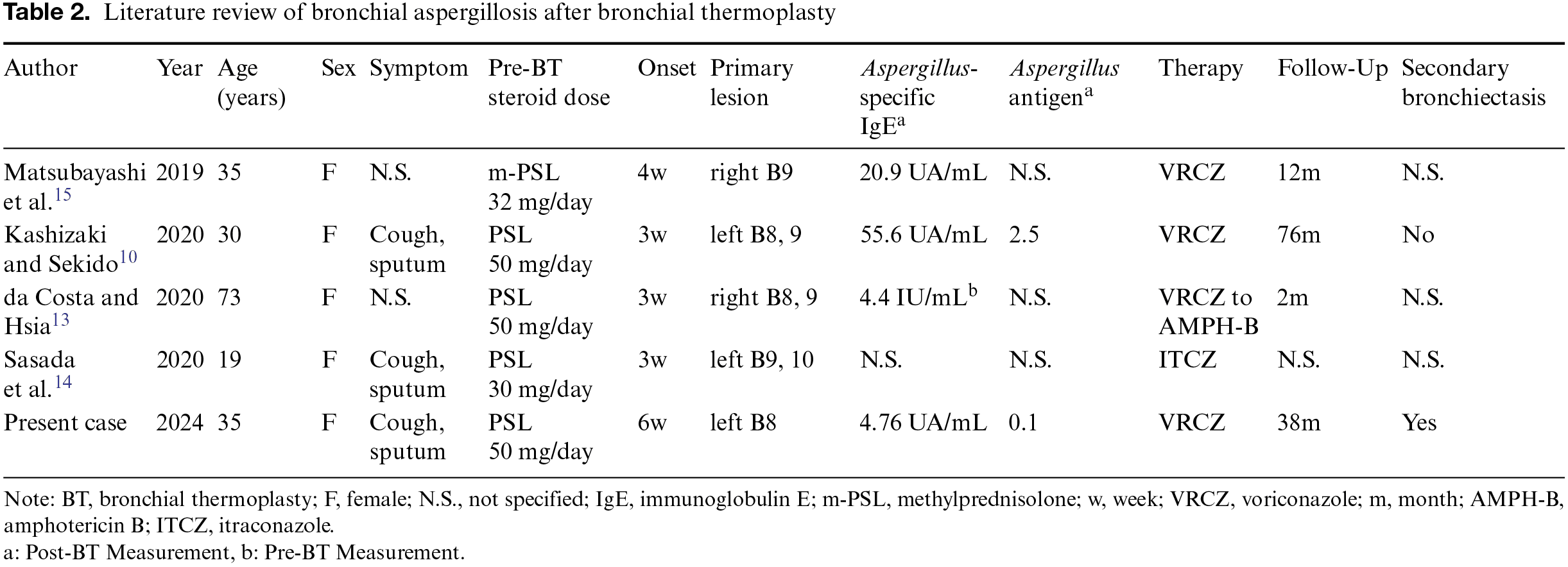
In a prior report, a six-year follow-up of a patient treated for aspergillosis revealed only mild bronchiectasis on CT, with no significant increase in sputum production.10 In contrast, the present case showed persistent localized bronchiectasis and purulent sputum following bronchial aspergillosis. Additionally, a chronic lower respiratory tract infection caused by staphylococci emerged during the disease course, requiring ongoing treatment with erythromycin. This progression is similar to that of bronchiectasis secondary to ABPA in patients without a history of BT.21
Bronchiectasis is a chronic disease characterized by permanent dilation of the bronchi and various clinical manifestations, including cough and sputum production.4,22 In the EMBARC cohort, 61.9% of bronchiectasis cases were reported to be associated with secondary causes, with BA accounting for 6.9% and ABPA for 2.8%.4 Though it was once believed that bronchiectasis progressed continuously due to infection, recent research has introduced the widely accepted “vicious vortex” model.23 This model highlights the interplay of four factors contributing to disease progression, viz., chronic infection, lung destruction and dysfunction, neutrophilic inflammation, and impaired mucociliary clearance.5,23,24 The differences between the two cases we encountered suggest that the development of bronchiectasis may have been influenced by lung damage, impaired function, and chronic infection caused by BT, along with patient-specific factors such as neutrophilic inflammation and reduced mucociliary clearance.5,23
Advances in treatment strategies for bronchiectasis have improved management options. Long-term macrolide therapy has gained worldwide attention for managing frequent exacerbations unresponsive to short-term antimicrobial treatment. Studies such as EMBRACE,6 BLESS,8 and BAT7 have demonstrated the efficacy of macrolides in reducing exacerbations, particularly in patients with persistent P. aeruginosa infections. Additionally, meta-analyses have shown similar benefits in subgroups without P. aeruginosa infections,25 further supporting this approach. In fact, administering low-dose, long-term macrolide therapy reduced the frequency of exacerbations compared to pre-treatment levels. Recently, emerging therapies such as brensocatib, which targets neutrophil reprogramming by inhibiting dipeptidyl peptidase 1 (DPP1),9,26 have shown promise in improving outcomes for patients with bronchiectasis. These advancements provide hope for more effective management strategies in this challenging condition.
Conclusions
Bronchial aspergillosis, a rare complication following BT, is typically treated with antifungal agents; however, long-term follow-up is crucial as some cases may progress to bronchiectasis. While no standard treatment exists for these cases, recent strategies for managing bronchiectasis, such as long-term macrolide therapy and DPP1 inhibitors, may mitigate disease progression.
Acknowledgment
We appreciate the patient’s cooperation in the data collection.
Funding
None.
Author Contributions
F.K. and Y.S. were responsible for data curation. F.K. interpreted the data and drafted the original manuscript. Both authors have read the manuscript and agree with the content, data, and the revision process.
Data Availability Statement
The datasets used in the current study are available from the corresponding author upon reasonable request.
Ethical Statement
The article does not involve the participation of any animals. The patient provided written informed consent for the publication of this report and accompanying images.
Conflicts of Interest
The authors declare no conflicts of interest related to this study.
Supplemental Information
Supplemental information for this article can be found online at https://sup.jclinque.com/api/articles/57/download-suppl.
References
[1] Konishi K, Takeuchi K, Hayakawa N, et al. Biomarkers identifying the effectiveness of biologic agents in moderate to severe asthma: a systematic review and meta-analysis. J Clin Quest. October 15, 2024;1(2):31–41. doi:10.69854/jcq.2024.0005.
[2] Kashizaki F, Konishi K, Chen H, et al. Pretreatment asthma control test score as a predictive score for clinical remission after bronchial thermoplasty in younger patients with severe asthma and preserved lung function. J Asthma. July 2024;61(7):663–670. doi:10.1080/02770903.2023.2297375.
[3] Chen X, Zhi H, Wang X, et al. Efficacy of biologics in patients with allergic bronchopulmonary aspergillosis: a systematic review and meta-analysis. Lung. August 2024;202(4):367–383. doi:10.1007/s00408-024-00717-y.
[4] Chalmers JD, Polverino E, Crichton ML, et al. Bronchiectasis in Europe: data on disease characteristics from the European Bronchiectasis registry (EMBARC). Lancet Respir Med. July 2023;11(7):637–649. doi:10.1016/S2213-2600(23)00093-0.
[5] Choi H, McShane PJ, Aliberti S, et al. Bronchiectasis management in adults: state of the art and future directions. Eur Respir J. June 2024;63(6):2400518. doi:10.1183/13993003.00518-2024.
[6] Wong C, Jayaram L, Karalus N, et al. Azithromycin for prevention of exacerbations in non-cystic fibrosis bronchiectasis (EMBRACE): a randomised, double-blind, placebo-controlled trial. Lancet. August 18, 2012;380(9842):660–667. doi:10.1016/S0140-6736(12)60953-2.
[7] Altenburg J, de Graaff CS, Stienstra Y, et al. Effect of azithromycin maintenance treatment on infectious exacerbations among patients with non-cystic fibrosis bronchiectasis: the BAT randomized controlled trial. JAMA. March 27, 2013;309(12):1251–1259. doi:10.1001/jama.2013.1937.
[8] Serisier DJ, Martin ML, McGuckin MA, et al. Effect of long-term, low-dose erythromycin on pulmonary exacerbations among patients with non-cystic fibrosis bronchiectasis: the BLESS randomized controlled trial. JAMA. March 27, 2013;309(12):1260–1267. doi:10.1001/jama.2013.2290.
[9] Chalmers JD, Shteinberg M, Mall MA, et al. Cathepsin C (dipeptidyl peptidase 1) inhibition in adults with bronchiectasis: AIRLEAF, a phase II randomised, double-blind, placebo-controlled, dose-finding study. Eur Respir J. 2025;65(1):2401551. doi:10.1183/13993003.01551-2024.
[10] Kashizaki F, Sekido Y. Bronchial aspergillosis following bronchial thermoplasty. QJM. December 1, 2020;113(12):900. doi:10.1093/qjmed/hcaa101.
[11] Chaudhuri R, Rubin A, Sumino K, et al. Safety and effectiveness of bronchial thermoplasty after 10 years in patients with persistent asthma (BT10+): a follow-up of three randomised controlled trials. Lancet Respir Med. May 2021;9(5):457–466. doi:10.1016/S2213-2600(20)30408-2.
[12] Kashizaki F, Sekido Y. Response by Kashizaki to letter regarding article, ‘Bronchial aspergillosis following bronchial thermoplasty’. QJM. February 18, 2021;114(1):65. doi:10.1093/qjmed/hcaa116.
[13] da Costa DJ, Hsia DW. Aspergillus tracheobronchitis complicating bronchial thermoplasty. J Bronchol Interv Pulmonol. January 2020;27(1):68–71. doi:10.1097/LBR.0000000000000628.
[14] Sasada S, Ohmura K, Oguri T, et al. A case report of aspergillosis accompanied by saccular bronchodilation after bronchial thermoplasty in a 19-year-old woman. BMC Pulm Med. November 26, 2020;20(1):312. doi:10.1186/s12890-020-01352-y.
[15] Matsubayashi S, Iikura M, Numata T, et al. A case of Aspergillus and Nocardia infections after bronchial thermoplasty. Respirol Case Rep. February 2019;7(2):e00392. doi:10.1002/rcr2.392.
[16] Qiu M, Lai Z, Wei S, et al. Bronchiectasis after bronchial thermoplasty. J Thorac Dis. October 2018;10(10):E721–E726. doi:10.21037/jtd.2018.09.116.
[17] Asano K, Hebisawa A, Ishiguro T, et al. New clinical diagnostic criteria for allergic bronchopulmonary aspergillosis/mycosis and its validation. J Allergy Clin Immunol. April 2021;147(4):1261–1268.e5. doi:10.1016/j.jaci.2020.08.029.
[18] Sehgal IS, Choudhary H, Dhooria S, et al. Is there an overlap in immune response between allergic bronchopulmonary and chronic pulmonary aspergillosis? J Allergy Clin Immunol Pract. March 2019;7(3):969–974. doi:10.1016/j.jaip.2018.08.034.
[19] Ito Y, Takazono T, Obase Y, et al. Serum cytokines usefulness for understanding the pathology in allergic bronchopulmonary aspergillosis and chronic pulmonary aspergillosis. J Fungi (Basel). April 23, 2022;8(5). doi:10.3390/jof8050436.
[20] Ishiguro T, Isono T, Maruyama T, et al. Overlap of chronic pulmonary aspergillosis on allergic bronchopulmonary aspergillosis. Intern Med. August 2024;1(15):2167–2171. doi:10.2169/internalmedicine.2562-23.
[21] Nigro M, Laska IF, Traversi L, et al. Epidemiology of bronchiectasis. Eur Respir Rev. October 2024;33(174). doi:10.1183/16000617.0091-2024.
[22] Polverino E, Goeminne PC, McDonnell MJ, et al. European respiratory society guidelines for the management of adult bronchiectasis. Eur Respir J. 2017;50(3):1700629. doi:10.1183/13993003.00629-2017.
[23] Chalmers JD, Mall MA, Chotirmall SH, et al. Targeting neutrophil serine proteases in bronchiectasis. Eur Respir J. 2025;65(1):2401050. doi:10.1183/13993003.01050-2024.
[24] Giam YH, Shoemark A, Chalmers JD. Neutrophil dysfunction in bronchiectasis: an emerging role for immunometabolism. Eur Respir J. 2021;58(2):2003157. doi:10.1183/13993003.03157-2020.
[25] Chalmers JD, Boersma W, Lonergan M, et al. Long-term macrolide antibiotics for the treatment of bronchiectasis in adults: an individual participant data meta-analysis. Lancet Respir Med. October 2019;7(10):845–854. doi:10.1016/S2213-2600(19)30191-2.
[26] Chalmers JD, Moffitt KL, Suarez-Cuartin G, et al. Neutrophil Elastase activity is associated with exacerbations and lung function decline in bronchiectasis. Am J Respir Crit Care Med. May 15, 2017;195(10):1384–1393. doi:10.1164/rccm.201605-1027OC.
 |
Copyright: © 2025 Kashizaki and Sekido. This work is licensed under a Creative Commons Attribution 4.0 International License, which permits unrestricted use, distribution, and reproduction in any medium, provided the original work is properly cited. |