| Journal of Clinical Question. 2026; 3(1): e104; https://doi.org/10.69854/jcq.2026.0006 Advance access publication date 13 February 2026 |
 |
Case Report
Infected Abdominal Aortic Aneurysm with Variable Clinical Presentations: A Case Series
Zaiqiang Yu *, Norihiro Kondo, Masahito Minakawa
Department of Thoracic and Cardiovascular Surgery, Hirosaki University Graduate School of Medicine.
*Corresponding Author: e-mail: yuzaiqiang@hirosaki-u.ac.jp
Submitted: January 22, 2026 Accepted: February 12, 2026
Clinical Question Box
What is the optimal diagnostic and therapeutic approach for infected abdominal aortic aneurysms?
Infected abdominal aortic aneurysms present with variable clinical features and may have negative blood cultures. Contrast-enhanced computed tomography is essential for diagnosis. Definitive treatment requires antimicrobial therapy and surgical excision of infected tissue, with open repair providing reliable source control. Adjunctive measures such as antibiotic-treated grafts and omental implantation may reduce reinfection risk, and postoperative antibiotics should be tailored to clinical and laboratory findings.
Abstract
Infected abdominal aortic aneurysm (AAA) is a rare but life-threatening condition requiring prompt diagnosis and aggressive management. We report three cases of infected infrarenal AAA treated with open surgical repair. The patients, aged 47–79 years, presented with variable clinical features, including localized pain, systemic inflammatory response, and rapid aneurysmal enlargement. Blood cultures were positive in two cases, while one case showed negative cultures despite clear radiologic and intraoperative evidence of infection. Contrast-enhanced computed tomography was crucial for diagnosis, revealing irregular aneurysm morphology, intramural gas, marked aortic wall enhancement, or rapid expansion. All patients underwent complete aneurysm resection with prosthetic Y-graft replacement using locally treated grafts, omental implantation, and perioperative antimicrobial therapy. One patient underwent endovascular aneurysm repair as a bridging procedure prior to definitive open surgery. No perioperative mortality, graft infection, or major complications occurred. These cases highlight the importance of open surgical repair with aggressive source control, adjunctive local anti-infective measures, and appropriate antimicrobial therapy to achieve favorable early outcomes in infected AAA.
Keywords: Infected abdominal aortic aneurysm, open surgery, treatment strategy, case report
Introduction
Infected abdominal aortic aneurysm (AAA), often categorized as an infective or mycotic aortic aneurysm,1 is a rare but highly lethal condition, accounting for approximately 0.7%–3% of all aortic aneurysms.2 Despite its low incidence, reported mortality remains substantial from 21% to 44%, and infected AAA contributes disproportionately to urgent surgical admissions and intensive inpatient care.3,4 Definitive management requires prompt antimicrobial therapy and surgical source control with excision of infected tissue and arterial reconstruction.5 Although endovascular approaches are increasingly used, open repair remains essential in cases of extensive infection, despite ongoing controversy regarding prosthetic reconstruction in contaminated fields. Adjunctive strategies, including antibiotic-treated grafts and coverage with well-vascularized tissue such as omental implantation, are commonly employed to reduce reinfection risk.6 Here, we report three cases of infected AAA treated with open revascularization, complete aneurysm resection, prosthetic graft reconstruction with local anti-infective treatment, omental implantation, and perioperative antimicrobial therapy.
Case Presentation
Case 1
A 47-year-old man presented with left knee pain and swelling without fever and initially visited a local hospital. Laboratory evaluation revealed marked inflammatory responses, with a white blood cell (WBC) count of 22,590/μL and C-reactive protein (CRP) level of 20.57 mg/dL. Renal function was preserved with a serum creatinine of 0.97 mg/dL. Blood cultures were positive for Enterococcus faecalis. Contrast-enhanced computed tomography (CT) demonstrated a 25-mm AAA with an irregular morphology (Figs. 1A and 1B), raising suspicion for an infected AAA. The patient was therefore referred to our institution.
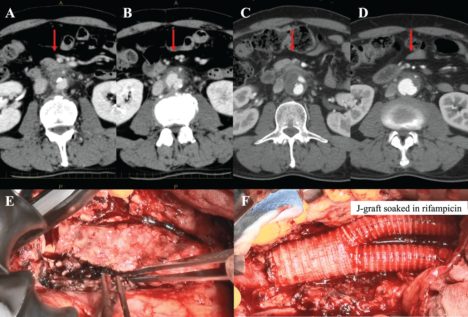
Figure 1. Infected abdominal aortic aneurysm with rupture. (A, B) Contrast-enhanced computed tomography (CT) showed a 25-mm abdominal aortic aneurysm with an irregular shape. (C, D) Follow-up CT after 2 weeks of antibiotic therapy showed enlargement of the aneurysm to 30 mm with suspected rupture into the inferior vena cava. (E, F) Intraoperative findings showed Y-graft replacement with aneurysm resection and omental implantation.
Antimicrobial therapy was initiated and continued for 2 weeks, during which no recurrence of fever was observed. However, follow-up CT showed aneurysm enlargement to 30 mm with evidence of impending rupture into the inferior vena cava (Figs. 1C and 1D). The patient subsequently underwent open Y-graft replacement with complete aneurysm resection and omental implantation (Figs. 1E and 1F). A prosthetic graft (J-Graft 18/11/11 mm; Japan Lifeline Co., Ltd., Tokyo, Japan) was soaked in rifampicin for 30 min prior to implantation. Although cultures of the aneurysm wall were negative, antimicrobial therapy was continued for 4 weeks postoperatively. The postoperative course was uneventful, and the patient was discharged without complications.
Case 2
A 79-year-old man presented with generalized body pain of 1 week’s duration. Laboratory testing revealed severe inflammatory responses, with a WBC count of 29,850/μL and CRP level of 44.74 mg/dL, along with impaired renal function with serum creatinine of 1.70 mg/dL. Blood cultures were positive for Staphylococcus pyogenes. Contrast-enhanced CT identified a 64 × 58-mm infrarenal AAA containing intramural gas, suggestive of infection (Figs. 2A and 2B).
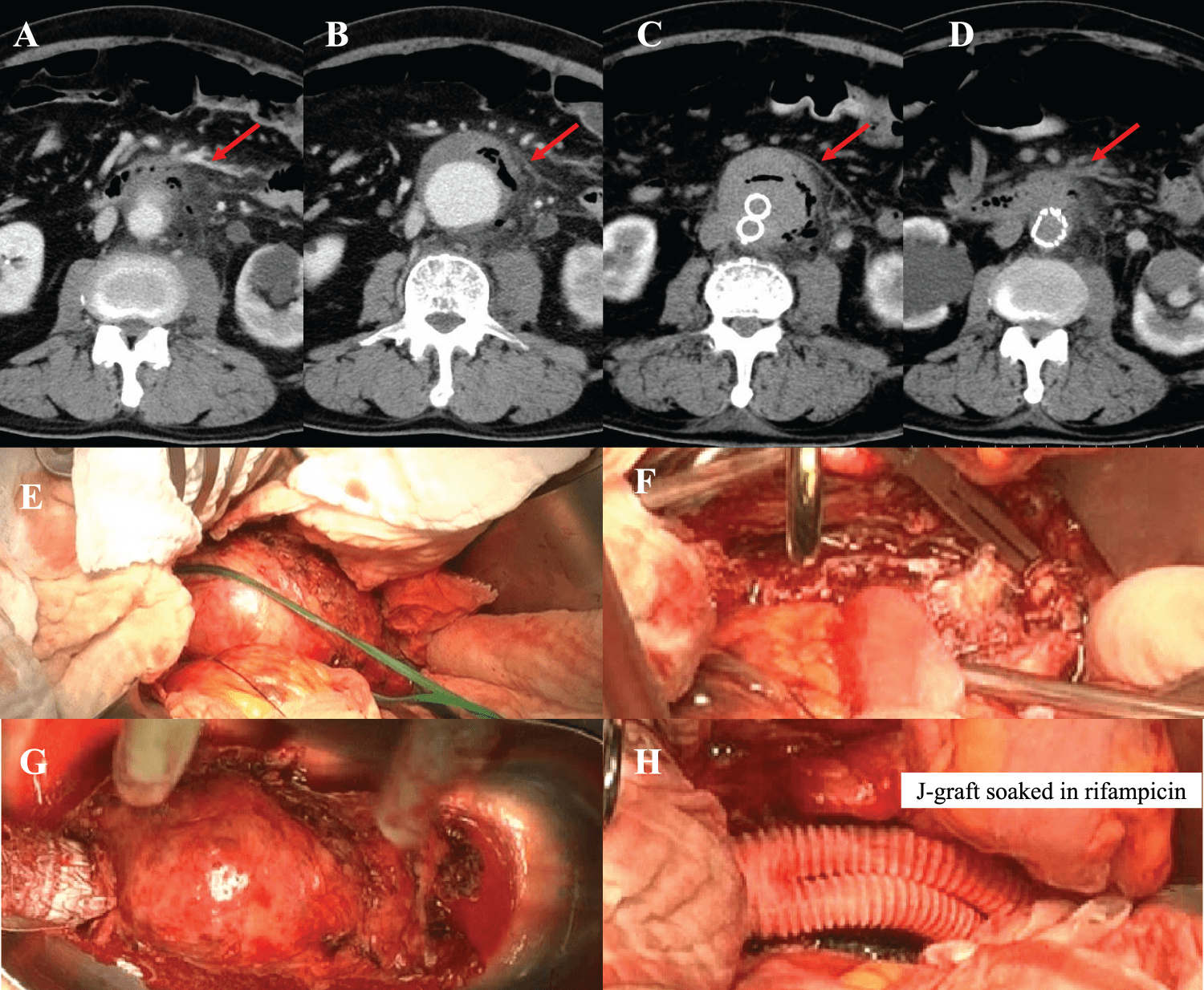
Figure 2. Infected abdominal aortic aneurysm treated with endovascular aneurysm repair (EVAR) and open repair. (A, B) Contrast-enhanced computed tomography (CT) showed a 64 × 58-mm infrarenal abdominal aortic aneurysm with intramural air. (C, D) Post-EVAR imaging showed successful exclusion of the aneurysm. (E–H) Intraoperative findings showed Y-graft replacement and omental implantation using a rifampicin-soaked graft.
Endovascular aneurysm repair (EVAR) was initially performed to prevent imminent rupture, followed by continued antimicrobial therapy for 2 weeks (Figs. 2C and 2D). Inflammatory markers improved substantially (WBC 8,040/μL; CRP 6.13 mg/dL). Definitive open surgery was subsequently undertaken, consisting of Y-graft replacement with complete aneurysm resection and omental implantation (Figs. 2E–2H). A rifampicin-soaked prosthetic graft (J-Graft 18/9/9 mm; Japan Lifeline Co., Ltd.) was used. Intraoperatively, a large amount of purulent material was observed, and cultures from the aneurysm wall were positive. Antimicrobial therapy was continued for 12 days postoperatively. The patient’s postoperative course was favorable, with no complications.
Case 3
A 69-year-old man with a history of coronary artery bypass grafting (left internal thoracic artery to the left anterior descending artery; right gastroepiploic artery to the posterior descending and atrioventricular branches) 2 years earlier presented with sudden loss of consciousness and fever of 37.8°C. Contrast-enhanced CT revealed rapid enlargement of an infrarenal AAA from 31 to 62 mm with marked aortic wall enhancement (Figs. 3A–3C). Laboratory findings showed elevated inflammatory markers (WBC 14,610/μL; CRP 16.05 mg/dL) with preserved renal function (serum creatinine 1.03 mg/dL). Blood cultures were negative.
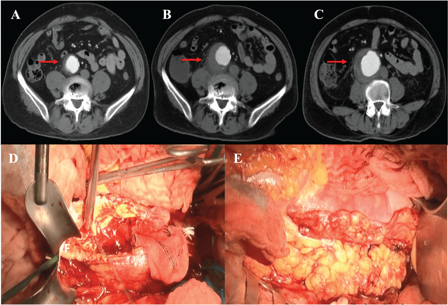
Figure 3. Acute enlargement of infected abdominal aortic aneurysm. (A–C) Contrast-enhanced computed tomography (CT) showed rapid enlargement of the abdominal aortic aneurysm from 31 to 62 mm with marked aortic wall enhancement. (D, E) Operative findings showed Y-graft replacement with aneurysm resection and omental implantation using a pyoctanine-treated graft.
Given the rapid aneurysm expansion and high rupture risk, emergent open repair was performed with complete aneurysm resection, Y-graft replacement, and omental implantation (Figs. 3D and 3E). A pyoctanine-treated prosthetic graft (J-Graft 18/9/9 mm; Japan Lifeline Co., Ltd.) was used. Cultures from the aneurysm wall were positive, and postoperative antimicrobial therapy was administered 2 two weeks. The postoperative course was uneventful, and the patient was discharged without complications. The patient was followed up approximately every 2 months, and annually thereafter, as with previous patients.
Discussion
Current recommendations for the diagnosis and management of aortic infections are largely based on expert consensus, as high-quality randomized evidence is limited. Widely used frameworks and guidelines, including the MAGIC criteria and the European Society for Vascular Surgery guidelines,7,8 were developed through structured consensus methods such as the Delphi process, providing practical guidance in areas where robust clinical trials are not feasible. The present case series highlights the heterogeneous clinical presentations, microbiological findings, and surgical strategies associated with infected AAA, while reinforcing the importance of aggressive source control combined with appropriate antimicrobial therapy. Consistent with previous reports, the three patients in this series demonstrated variable clinical features, ranging from localized pain without fever to systemic inflammatory response and hemodynamic instability.9,10 Notably, blood cultures were positive in two cases but negative in one, underscoring that negative blood cultures do not exclude the diagnosis of infected AAA.11
In all cases, contrast-enhanced CT played a central role in diagnosis by demonstrating characteristic findings such as irregular aneurysm morphology, rapid aneurysmal enlargement, intramural gas, and marked aortic wall enhancement.12 These imaging features, in conjunction with inflammatory markers and clinical presentation, were critical in guiding timely intervention.13 FDG-PET/CT is increasingly recognized as a useful adjunctive imaging modality in the diagnosis of infected aortic aneurysms and aortic graft infections.14 By detecting metabolically active inflammatory or infectious tissue, it can improve diagnostic confidence when conventional contrast-enhanced CT findings are equivocal, and it is included as an imaging criterion within the MAGIC framework. However, despite its clinical value, FDG-PET/CT for suspected aortic infection is generally not covered by the Japanese national health insurance system, except for limited indications such as confirmed large-vessel vasculitis, which may restrict its routine use in clinical practice.
All patients in this series ultimately underwent open surgical repair with complete aneurysm resection, reflecting the continued importance of open surgery in cases of extensive infection or high rupture risk. Although EVAR has been increasingly utilized as a less invasive option, concerns remain regarding persistent infection due to incomplete source control.8 In Case 2, EVAR was employed as a bridge to surgery to stabilize the patient and reduce rupture risk, allowing optimization of inflammatory status prior to definitive open repair. In general, EVAR was selected as a bridge strategy for patients in poor overall condition and at high risk of rupture, for whom immediate open repair was considered too hazardous.
Reconstruction in an infected field remains controversial, particularly when prosthetic grafts are used. In this series, all patients underwent prosthetic graft replacement with adjunctive local anti-infective measures, including rifampicin- or pyoctanine-treated grafts and omental implantation.15 These strategies aim to reduce bacterial colonization and enhance local immune defense. Omental implantation, in particular, provides well-vascularized tissue coverage and has been widely adopted to decrease the risk of graft infection and reinfection.16 The absence of postoperative infectious complications in all three patients supports the effectiveness of this combined approach.
The duration of antimicrobial therapy varied among the cases, reflecting differences in microbiological findings and clinical response. Although no consensus exists regarding the optimal duration of postoperative antibiotics, continued therapy tailored to culture results and inflammatory markers remains essential.17 When complete excision of infected tissue was achieved and postoperative laboratory markers indicated adequate infection control, antibiotics were generally discontinued after approximately 2 weeks. Notably, all patients demonstrated favorable early outcomes, with no perioperative mortality or major complications.
This case series is limited by its small sample size and short-term follow-up; however, it illustrates practical decision-making in real-world clinical settings. Our experience suggests that prompt recognition, aggressive surgical source control, use of locally treated prosthetic grafts, omental coverage, and individualized antimicrobial therapy can result in satisfactory early outcomes, even in complex and high-risk presentations.
Conclusion
Infected AAA is a rare and life-threatening condition requiring prompt diagnosis and aggressive treatment. Open surgical repair with complete aneurysm resection, adjunctive local anti-infective measures, omental implantation, and appropriate antimicrobial therapy can achieve favorable early outcomes.
Acknowledgment
We appreciate the patients’ cooperation in data collection.
Funding Source
None.
Author Contributions
Z.Y. was responsible for data curation, data interpretation, and drafting of the original manuscript. N.K. and M.M. were responsible for the revision. All authors have read and approved the manuscript and agree with the content and data.
Data Availability Statement
The datasets used in the current study are available from the corresponding author upon reasonable request.
Generative AI Declaration
During the preparation of this manuscript, the authors used OpenAI to assist with proofreading. All content was subsequently reviewed and edited by the authors, who assume full responsibility for the accuracy and integrity of the published work.
Ethical Statement
This study did not involve the participation of any animals. Informed consent was obtained from all patients. Institutional Review Board approval was waived because this was a case report.
Conflict of Interest
The authors report no conflicts of interest in this work.
Supplemental Information
Supplemental information for this article can be found online at https://sup.jclinque.com/api/articles/104/download-suppl.
References
[1] Wu S, Yan J, Kang Z, Zhang J. Clinical and computed tomography angiography characteristics of infected vs. non-infected abdominal aortic aneurysm: a comparative study. Abdom Radiol (NY). December 2024;49(12):4334–4340. doi:10.1007/s00261-024-04312-w.
[2] Huang YK, Chen CL, Lu MS, et al. Clinical, microbiologic, and outcome analysis of mycotic aortic aneurysm: the role of endovascular repair. Surg Infect (Larchmt). June 2014;15(3):290–298. doi:10.1089/sur.2013.011.
[3] Shinya N, Seki M, Karaushi H, Asakura T, Yoshitake A, Mitsutake K. Infective native aortic and iliac artery aneurysms: clinical profiles and short-term outcomes from a single-center cohort. J Infect Chemother. 2025;31(4):102644. doi:10.1016/j.jiac.2025.102644.
[4] Premnath S, Zaver V, Hostalery A, Rowlands T, Quarmby J, Singh S. Mycotic abdominal aortic aneurysms–A tertiary centre experience and formulation of a management protocol. Ann Vasc Surg. July 1, 2021;74: 246–257. doi:10.1016/j.avsg.2020.12.025.
[5] Hirano K, Tokui T, Nakamura B, et al. Hybrid therapy for mycotic aortic aneurysm with stent-graft and video-assisted thoracoscopic debridement. Ann Vasc Dis. March 25, 2019;25(1):69–73. doi:10.3400/avd.cr.18-00119.
[6] Caradu C, Jolivet B, Puges M, Cazanave C, Ducasse E, Berard X. Reconstruction of primary and secondary aortic infections with an antimicrobial graft. J Vasc Surg. 2023;77(4):1226–1237.e10. doi:10.1016/j.jvs.2022.11.065.
[7] Lyons OT, Baguneid M, Barwick TD, et al. Diagnosis of aortic graft infection: a case definition by the management of aortic graft infection collaboration (MAGIC). Eur J Vasc Endovasc Surg. December 2016;52(6):758–763. doi:10.1016/j.ejvs.2016.09.007.
[8] Committee EG, Document R, Wanhainen A, et al. Editor’s choice-European society for vascular surgery 2024 clinical practice guidelines on the management of abdominal aorto-iliac artery aneurysms. Eur J Vasc Endovasc Surg. 2024;67(2):192–331. doi:10.1016/j.ejvs.2023.11.002.
[9] Matsui K, Takahashi K, Tashiro M, et al. Clinical and microbiological characteristics and challenges in diagnosing infected aneurysm: a retrospective observational study from a single center in Japan. BMC Infect Dis. June 30, 2022;22(1):585. doi:10.1186/s12879-022-07567-0.
[10] Gonzalez-Urquijo M, Salgado-Garza G, Martin AM, et al. Infective native visceral artery aneurysm (INVAA): a systematic review of etiology, treatment, and outcomes. Vasc Med. August 1, 2025;30(4510–521. doi:10.1177/1358863X251326537.
[11] Dang Q, Statius van Eps RG, Wever JJ, et al. Nationwide study of the treatment of mycotic abdominal aortic aneurysms comparing open and endovascular repair in The Netherlands. J Vasc Surg. August 1, 2020;72(2):531–540. doi:10.1016/j.jvs.2019.09.060.
[12] Zhang N, Xiong W, Li Y, et al. Imaging features of mycotic aortic aneurysms. Quant Imaging Med Surg. June 2021;11(6):2861–2878. doi:10.21037/qims-20-941.
[13] Jutidamrongphan W, Kritpracha B, Sörelius K, Hongsakul K, Suwannanon R. Features of infective native aortic aneurysms on computed tomography. Insig into Imag. January 8, 2022;13(1):2. doi:10.1186/s13244-021-01135-x.
[14] Badarna M, Keidar Z, Arnon-Sheleg E. FDG PET/CT in vascular graft infection: a pictorial review. Q J Nucl Med Mol Imaging. March 2025;69(1):61–68. doi:10.23736/s1824-4785.25.03612-x.
[15] Colacchio EC, D’Oria M, Grando B, et al. A systematic review of in-situ aortic reconstructions for abdominal aortic graft and endograft infections: outcomes of currently available options for surgical replacement. Ann Vasc Surg. 2023;95:307–316. doi:10.1016/j.avsg.2023.03.005.
[16] Yamanaka K, Okada K, Kato D, et al. Long-term outcomes and evolving trends in thoracic aortic infections: a 25-year, single-center study in Japan. JTCVS Open. December 2025;28:1–12. doi:10.1016/j.xjon.2025.10.004.
[17] Wu S-J, Sun S, Tan Y-H, Chien C-Y. Analysis of antibiotic strategies to prevent vascular graft or endograft infection after surgical treatment for infective native aortic aneurysms: a systematic review. Antimicrob Resist Infect Cont. October 1, 2024;13(1):116. doi:10.1186/s13756-024-01477-3.
 |
Copyright: © 2026 Yu et al. This work is licensed under a Creative Commons Attribution 4.0 International License, which permits unrestricted use, distribution, and reproduction in any medium, provided the original work is properly cited. |