| Journal of Clinical Question. 2026; 3(1): e103 https://doi.org/10.69854/jcq.2025.0040 Advance access publication date 31 January 2026 |
 |
Case Report
Diagnostic Challenges in Immune-Mediated Thrombotic Thrombocytopenic Purpura: A Case Report of Initial Misdiagnosis
California Northstate University College of Medicine, Elk Grove, CA, USA.
*Corresponding Author: e-mail: garychumd@gmail.com
Submitted: December 02, 2025 Accepted: January 30, 2026
Clinical Question Box
How should immune-mediated thrombotic thrombocytopenic purpura (iTTP) be identified and managed in patients with atypical presentations and multiple comorbidities?
iTTP should be suspected in patients with unexplained thrombocytopenia and hemolytic anemia, even without the classic pentad or schistocytes on initial peripheral smear, particularly in those with underlying autoimmune disease. Prompt initiation of therapeutic plasma exchange is warranted while awaiting confirmatory ADAMTS13 (a disintegrin and metalloproteinase with thrombospondin type 1 motif, member 13) testing, with adjunctive therapies such as corticosteroids and caplacizumab to reduce immune-mediated activity and thrombotic risk. Early recognition and timely treatment are crucial to minimizing morbidity and mortality, especially in patients with complex or confounding comorbidities.
Abstract
Thrombotic thrombocytopenic purpura (TTP) is a rare, life-threatening thrombotic microangiopathy caused by severe ADAMTS13 (a disintegrin and metalloproteinase with thrombospondin type 1 motif, member 13) deficiency. Diagnosis is often challenging because most patients do not present with the classic pentad of findings. We report a 56-year-old woman with multiple comorbidities, including systemic lupus erythematosus and antiphospholipid syndrome, who presented with neurologic symptoms, hemolytic anemia, and severe thrombocytopenia without initial schistocytes. Her condition was initially misattributed to isoniazid-induced thrombocytopenia, resulting in delayed recognition of TTP. Immune-mediated TTP was suspected based on her autoimmune disease history and was definitively confirmed when ADAMTS13 activity was <1%. Initiation of plasma exchange, corticosteroids, and caplacizumab led to rapid hematologic recovery. This case underscores the importance of maintaining a high index of suspicion for immune-mediated TTP, particularly in patients with underlying autoimmune disease, as early diagnosis and prompt treatment are critical to preventing fatal thrombotic complications.
Keywords: Thrombotic thrombocytopenic purpura, ADAMTS13, thrombotic microangiopathy, diagnostic challenge, plasma exchange, case report,
Introduction
Thrombotic thrombocytopenic purpura (TTP) is a rare, life-threatening autoimmune thrombotic microangiopathy with an estimated incidence of 1.5–6 cases per million adults annually.1 TTP occurs in two distinct forms. The inherited variant arises from pathogenic mutations in the ADAMTS13 (a disintegrin and metalloproteinase with thrombospondin type 1 motif, member 13) gene,2 whereas the acquired variant, known as immune-mediated TTP (iTTP), develops due to autoantibodies that impair ADAMTS13 function.3 Although TTP is classically described by a pentad of thrombocytopenia, microangiopathic hemolytic anemia, neurologic abnormalities, renal dysfunction, and fever, most patients present with only a subset of these features, contributing to diagnostic uncertainty.4
In contemporary clinical practice, the absence of the full pentad and the nonspecific nature of early symptoms, such as fatigue, altered mental status, abdominal pain, or renal impairment, often lead to initial misdiagnosis as immune thrombocytopenic purpura, hemolytic uremic syndrome, sepsis, or disseminated intravascular coagulation.5 Diagnostic delays may be further compounded by atypical laboratory findings, including the absence of schistocytes on peripheral blood smear or ADAMTS13 activity levels initially inconsistent with TTP. These challenges are exemplified in the present case of a 56-year-old woman whose condition was initially attributed to isoniazid (INH)-induced thrombocytopenia, resulting in delayed diagnostic clarity and definitive treatment.
Case Report
A 56-year-old woman presented with dizziness. Her medical history was notable for systemic lupus erythematosus (SLE), antiphospholipid syndrome (APS), hypertension, hyperlipidemia, obesity, nonalcoholic fatty liver disease, type 2 diabetes mellitus, hypothyroidism, and latent tuberculosis (TB). One week prior, she had been hospitalized for abdominal pain and jaundice. At that time, laboratory evaluation revealed thrombocytopenia and hemolytic anemia, which were attributed to INH therapy for latent TB for 2 months (Fig. 1). She received blood transfusions, INH was discontinued, and she was discharged with plans for outpatient laboratory monitoring.
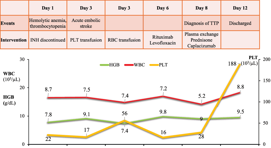
Figure 1. Clinical course and laboratory trends. HGB, hemoglobin; RBC, red blood cell; PLT, platelet; WBC, white blood cell; TTP, thrombotic thrombocytopenic purpura; INH, isoniazid.
While awaiting follow-up bloodwork, the patient experienced acute-onset dizziness accompanied by decreased peripheral vision, anxiety, and chest discomfort on Day 3. A stroke alert was activated in the emergency department after examination revealed facial droop, mild dysarthria, tongue paresthesia, and anomia. Computed tomography and magnetic resonance imaging demonstrated embolic infarcts involving the left cerebellum, right occipital lobe, and right posterior temporal lobe, without evidence of large-vessel occlusion. Multifocal bilateral cerebral and cerebellar infarctions without identifiable responsible vessels suggest a microvascular thrombotic process rather than an embolic stroke. The neurologic deficits were classified as mild and nondisabling, consistent with an acute embolic stroke.
Laboratory evaluation at that time showed Coombs-negative hemolytic anemia with a hemoglobin of 9.1 g/dL and severe thrombocytopenia with a platelet count of 17,000/µL. Fibrinogen levels were elevated, and the initial peripheral blood smear was negative for schistocytes. One unit of platelets was transfused. The findings were interpreted as consistent with thrombocytopenia due to platelet consumption rather than impaired production. Considering the pathophysiology of Coombs-negative hemolytic anemia, INH-induced bicytopenia was deemed unlikely. However, continued downtrending hemoglobin and platelet levels prompted transfusion of two units of packed red blood cells and an additional unit of platelets later that day.
Despite supportive management, the patient’s anemia and thrombocytopenia persisted and were initially attributed to medication effects and hepatic dysfunction on Day 6. A repeat peripheral blood smear demonstrated occasional schistocytes. iTTP was considered while ADAMTS13 activity results were still pending. APS was also included in the differential diagnosis; however, laboratory findings, including Coombs-negative hemolysis, made this diagnosis less likely. Given her recent embolic stroke and ongoing severe thrombocytopenia, rituximab therapy was initiated. Concern for TB reactivation prompted consultation with infectious disease specialists, and levofloxacin was started for latent TB treatment.
By Day 8, laboratory studies showed gradual improvement in hemolytic anemia; however, thrombocytopenia persisted. ADAMTS13 activity returned at <1%, confirming the diagnosis of TTP. In the context of the patient’s history of SLE and APS, iTTP was considered the most likely diagnosis. The patient was started on daily plasma exchange, prednisone at 1 mg/kg, and caplacizumab. She demonstrated a rapid hematologic response, with platelet counts normalizing to 188,000/µL after 4 days of therapy.
The patient was discharged on Day 12 with instructions to continue prednisone and caplacizumab, with close outpatient hematology follow-up for ongoing TTP management and steroid tapering. She was also advised to continue levofloxacin for treatment of latent TB.
Discussion
TTP is a medical emergency, with acute cases reaching mortality rates of greater than 90% if left untreated and 15% with early suspicion and clinical intervention.6 Adverse outcomes include coronary thrombosis, leading to myocardial infarction, congestive heart failure, and death.7 In iTTP cases, microvascular thrombosis is more frequently observed.8 First-line treatment for an acute episode of TTP is immediate therapeutic plasmapheresis to improve levels of functioning ADAMTS13.9 Plasmapheresis is continued until platelet counts, lactate dehydrogenase levels, and organ function normalize.10 Adjunct therapies include corticosteroids, rituximab, caplacizumab, and splenectomy, all of which are considered in cases unresponsive to plasmapheresis alone.11
This case highlights the importance of a comprehensive approach to the management of TTP and the challenges faced when patients present with numerous overlapping, nonspecific comorbidities. TTP is a diagnosis that is usually considered after a patient exhibits clinical signs of suspicion, including a pentad of thrombocytopenia, renal dysfunction, hemolytic anemia, fever, and neurologic deficits. Despite this classic association, only about 10% of cases present with all five symptoms.12 While it is now widely accepted that suspicion for TTP must include only hemolytic anemia and thrombocytopenia, abnormal presentations can manifest without both criteria, as evident in this case.13 Schistocytes can be absent in initial presentations of TTP in up to 30% of patients.14 This case emphasizes that the absence of schistocytes on peripheral blood smear should not preclude consideration of TTP in the differential diagnosis, as early diagnosis and prompt treatment are essential for optimal outcomes. Raised clinical suspicion from other presenting symptoms must be considered and properly worked up to avoid overlook potentially fatal pathologies.
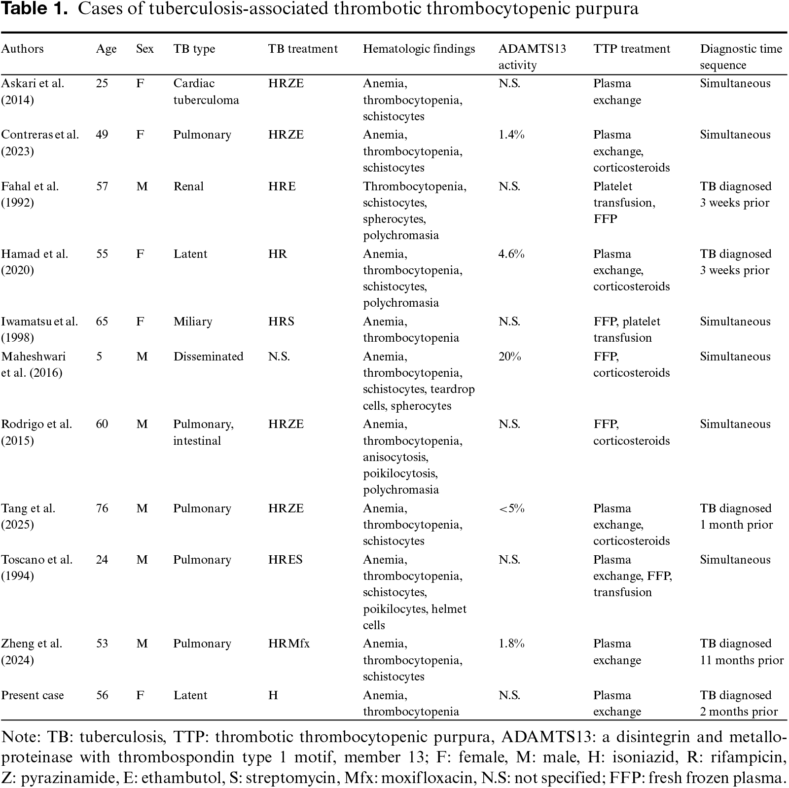
Whether an innate pathophysiologic connection between TB and TTP exists remains unknown. Previously published case reports describing concurrent TB and TTP were reviewed (Table 1).15–23 A total of 11 cases have been reported, including one pediatric case. The median age among adult patients was 56 years, with no apparent sex predilection. TB manifestations were heterogeneous, most commonly pulmonary, but also included latent, miliary, disseminated, renal, and cardiac forms. Nearly all patients presented with anemia, thrombocytopenia, and schistocytes on peripheral blood smear, consistent with TTP. When reported, ADAMTS13 activity was markedly reduced, supporting a diagnosis of immune-mediated TTP. Plasma exchange, often combined with corticosteroids, was the primary treatment modality, whereas earlier cases frequently relied on fresh frozen plasma and blood transfusions. In most cases, TB and TTP were diagnosed concurrently (54.5%, 6/11). Despite these observations, the temporal association between TB and TTP varied across cases, and a definitive causal relationship has not been established, suggesting that TB may act as a potential trigger in some patients, while in others the coexistence may be incidental.
Conclusion
Presentation of TTP can vary and manifest in ways not classically associated with it, emphasizing its importance as a clinically significant differential in the management of complex cases. Overlapping clinical symptoms of similar hemolytic pathologies alongside different multi-organ medical conditions can lead to difficult management and initial misdiagnosis of iTTP.
Acknowledgment
We appreciate the patient’s cooperation in data collection.
Funding Source
None.
Author Contributions
R.F. and K.A. were responsible for data curation, data interpretation, and drafting of the original manuscript. G.C. was responsible for the revision. All authors have read and approved the manuscript and agree with the content and data.
Data Availability Statement
The datasets used in the current study are available from the corresponding author upon reasonable request.
Generative AI Declaration
During the preparation of this manuscript, the authors used OpenAI to assist with proofreading. All content was subsequently reviewed and edited by the authors, who assume full responsibility for the accuracy and integrity of the published work.
Ethical Statement
The patient provided written informed consent for the publication of this case report. Identifying information has been omitted to protect patient confidentiality.
Conflict of Interest
The authors report no conflicts of interest in this work.
Supplemental Information
Supplemental information for this article can be found online at https://sup.jclinque.com/api/articles/103/download-suppl.
References
[1] Nuñez Zuno JA, Khaddour K. Thrombotic thrombocytopenic purpura evaluation and management. In: StatPearls. Treasure Island (FL): StatPearls Publishing; September 27, 2023.
[2] Stanley M, Killeen RB, Michalski JM. Thrombotic thrombocytopenic purpura. In: StatPearls. Treasure Island (FL): StatPearls Publishing; April 7, 2023.
[3] Murugan NL, Mensah R, Singh A. Raising suspicion of thrombotic thrombocytopenic purpura in a patient with a pre-existing autoimmune disease. Cureus. January 2025;17(1):e77623. doi:10.7759/cureus.77623.
[4] Issa L, Sandakly N, El Koubayati G, Khalil M, Haddad F. Renal involvement in thrombotic thrombocytopenic purpura: is it time to challenge the old paradigm?. Cureus. May 2024;16(5):e60259. doi:10.7759/cureus.60259.
[5] Scully M, Rayment R, Clark A, et al. A british society for haematology guideline: diagnosis and management of thrombotic thrombocytopenic purpura and thrombotic microangiopathies. Br J Haematol. November 2023;203(4):546–563. doi:10.1111/bjh.19026.
[6] Du P, Cristarella T, Goyer C, Moride Y. A systematic review of the epidemiology and disease burden of congenital and immune-mediated thrombotic thrombocytopenic purpura. J Blood Med. 2024;15:363–386. doi:10.2147/jbm.S464365.
[7] Brodsky MA, Sukumar S, Selvakumar S, et al. Major adverse cardiovascular events in survivors of immune-mediated thrombotic thrombocytopenic purpura. Am J Hematol. December 1, 2021;96(12):1587–1594. doi:10.1002/ajh.26341.
[8] Kayashima M, Sakai K, Harada K, et al. Strong association between insufficient plasma exchange and fatal outcomes in Japanese patients with immune-mediated thrombotic thrombocytopenic purpura. Int J Hematol. October 2021;114(4):415–423. doi:10.1007/s12185-021-03197-5.
[9] Zheng XL, Al-Housni Z, Cataland SR, et al. focused update of the 2020 ISTH guidelines for management of thrombotic thrombocytopenic purpura. J Thromb Haemost. November 2025;23(11):3711–3732. doi:10.1016/j.jtha.2025.06.002.
[10] Tran M-H, Jones J, Qiao J. Relapse and beyond: navigating the long-term clinical impacts of immune thrombotic thrombocytopenic purpura. Transfus Apheresis Sci. August 01, 2025;64(4):104174. doi:10.1016/j.transci.2025.104174.
[11] Lemiale V, Valade S, Mariotte E. Unresponsive thrombotic thrombocytopenic purpura (TTP): challenges and solutions. Ther Clin Risk Manag. 2021;17:577–587. doi:10.2147/tcrm.S205632.
[12] Matsumoto M, Miyakawa Y, Kokame K, et al. Diagnostic and treatment guidelines for thrombotic thrombocytopenic purpura (TTP) in Japan 2023. Int J Hematol. November 2023;118(5):529–546. doi:10.1007/s12185-023-03657-0.
[13] Smith L. Pathophysiology of thrombotic thrombocytopenia purpura. American Soc Clin Labor Sci. 2020. doi:10.29074/ascls.2020002261.
[14] Decker P, Moulinet T, Revuz S, Perez P, Jaussaud R. Thrombotic thrombocytopenic purpura without schistocytes: beware of misdiagnosis. Neurol Clin Pract. October 2021;11(5):e798–e800. doi:10.1212/cpj.0000000000001067.
[15] Askari R, Khouzam RN. Cardiac tuberculoma presenting as thrombotic thrombocytopenic purpura-hemolytic uremic syndrome. Heart Lung. March–April 2014;43(2):158–160. doi:10.1016/j.hrtlng.2013.11.001.
[16] Contreras K, Amorocho OMC, Giraldo JS. Acquired thrombotic thrombocytopenic purpura as a clinical manifestation of pulmonary tuberculosis: a case report. Germs. September 2023;13(3):259–265. doi:10.18683/germs.2023.1392.
[17] Fahal IH, Williams PS, Clark RE, Bell GM. Thrombotic thrombocytopenic purpura due to rifampicin. Bmj. April 4, 1992;304(6831):882. doi:10.1136/bmj.304.6831.882.
[18] Hamad H, Sahu KK, Dunn S, Milla L, Caffery A, Islam N. Rifampin induced thrombotic thrombocytopenic purpura. Indian J Hematol Blood Transfus. July 2020;36(3):575–577. doi:10.1007/s12288-019-01249-9.
[19] Iwamatsu H, Teramura T, Kikuchi M, Yoshida K. Case of chronic kidney failure with thrombotic thrombocytopenic purpura due to miliary tuberculosis. Nihon Naika Gakkai Zasshi. February 10, 1998;10(2):335–337.
[20] Maheshwari A, Bhaskar V, Mondal K, Seth A. Tuberculosis presenting as thrombotic thrombocytopenic purpura. Indian Pediat Case Rep. 2021;1(1):54–56. doi:10.4103/ipcares.ipcares_30_21.
[21] Rodrigo HF, Stavile RN, Correa M, Gutiérrez MM, Kicillof S, Boschero F. Thrombotic thrombocytopenic purpura and tuberculosis. A rare association. Medicina (B Aires). 2015;75(4):221–224. Púrpura trombótica trombocitopénica y tuberculosis. Una rara asociación.
[22] Toscano V, Bontadini A, Falsone G, et al. Thrombotic thrombocytopenic purpura associated with primary tuberculosis. Infection. January–February 1995;23(1):58–59. doi:10.1007/bf01710061.
[23] Zheng J, Pan X, Jiang Y. Pulmonary tuberculosis complicated by thrombotic thrombocytopenic purpura: a case report and literature review. Infect Drug Resist. 2024;17:3593–3598. doi:10.2147/idr.S477180.
 |
Copyright © 2026 Fan et al. This work is licensed under a Creative Commons Attribution 4.0 International License, which permits unrestricted use, distribution, and reproduction in any medium, provided the original work is properly cited. |