| Journal of Clinical Question. 2025; 2(3): e75 https://doi.org/10.69854/jcq.2025.0017 Advance access publication date 20 June 2025 |
 |
Case Report
Combined Respiratory and Heart Failure after Chlorine Exposure: A Rare Case Report
California Northstate University, College of Medicine.
*Corresponding Author: e-mail: shawncho32@gmail.com
Submitted: April 30, 2025 Accepted: June 19, 2025
Clinical Question Box
Can acute exposure to chlorine gas lead to cardiac dysfunction in addition to respiratory injury?
While chlorine gas primarily causes respiratory tract irritation and pulmonary injury, it can also lead to cardiac complications such as myocarditis or toxic cardiomyopathy, resulting in reduced left ventricular function. Clinicians should consider cardiac evaluation in patients presenting with chest symptoms or hemodynamic instability following chlorine exposure.
Abstract
Background: Chlorine gas is a common industrial chemical with known toxic effects on the respiratory system. However, cardiac complications are rarely reported. Case Presentation: We report the case of a 23-year-old previously healthy male who developed acute respiratory distress and reduced left ventricular systolic function following accidental exposure to chlorine gas. The patient presented with dyspnea, hypoxia, and chest discomfort. Imaging revealed bilateral infiltrates, and echocardiography showed a reduced left ventricular ejection fraction (40%–45%). He was diagnosed with heart failure with reduced ejection fraction, likely due to toxic cardiomyopathy or myocarditis. Supportive oxygen therapy and guideline-directed medical therapy for heart failure led to clinical improvement. Conclusion: This case highlights the potential for chlorine gas to cause not only pulmonary injury but also cardiac dysfunction. Clinicians should be aware of this rare but serious complication to enable timely diagnosis and management.
Keywords: Chlorine poisoning, heart failure, respiratory failure, case report,
Introduction
Chlorine gas (Cl2) is widely used in various industries, including plastics manufacturing, waste management, water treatment, and the pharmaceutical industry.1 Historically, it was employed as a chemical weapon during World War I.2 In modern times, most human exposures to chlorine are accidental, typically occurring in occupational settings, transportation-related incidents, or household accidents.3 At low concentrations (1–3 ppm), chlorine primarily irritates the eyes and oral mucous membranes, while concentrations around 15 ppm can also impact the pulmonary system. Prolonged exposure (30 minutes) to high concentrations, such as 430 ppm, can be fatal.4 Despite the potential for severe toxicity, most chlorine exposures tend to be nonfatal due to their accidental nature. Chlorine was identified as the most common inhalation irritant in the United States in 2016.5 The National Poison Data System reported 13,503 exposures to Cl2 in 2022 alone.6
Research into Cl2 toxicity remains limited, primarily due to the scarcity of controlled trials that quantify exposure levels and assess treatment options. Existing literature predominantly addresses pulmonary toxicity, while the cardiotoxic effects of Cl2 are not yet fully understood.7,8 Nonetheless, several reports suggest that Cl2 exposure may trigger serious cardiovascular events, such as atrial fibrillation, myocardial infarction, and acute ischemic stroke.9–11 Most studies have concentrated on the acute phase of these complications.12 Here, we present a case of a patient with no prior medical history who developed both respiratory and cardiac failure following Cl2 exposure.
Case Presentation
A 23-year-old man with a history of allergic rhinitis and obesity (body mass index: 44.1) presented to the emergency department (ED) with a 24-hour history of nonproductive cough, shortness of breath, and burning substernal chest pain, worsened by inspiration and physical exertion. The symptoms began after he was exposed for approximately five minutes to chlorine fumes while sleeping on a couch near a bucket of concentrated chlorine mixed with hot water, which his father had prepared to clean a pool. He reported eye and nasal irritation upon awakening. He was not on any medications, had no recent travel to tuberculosis (TB)-endemic areas, no known TB contact, and no history of smoking or baseline oxygen use. He denied fever, chills, nausea, vomiting, abdominal pain, or diarrhea.
Initial vital signs in the ED showed a blood pressure of 129/104 mmHg, heart rate of 116 bpm, temperature of 36.8°C, respiratory rate of 34 breaths per minute, and oxygen saturation of 84% on room air, which improved to 93% with 3 L/min of oxygen via nasal cannula (NC). The physical examination was notable for tachypnea and decreased breath sounds on bilateral lung auscultation. The complete blood count and complete metabolic panel were within normal limits. Viral swabs for coronavirus disease 2019, influenza A/B, and respiratory syncytial virus were negative. Laboratory findings included elevated serial troponin levels of 190 pg/mL (<26.2 pg/mL) and a brain natriuretic peptide level of 173 pg/mL (<18.4 pg/mL). Electrocardiogram showed sinus tachycardia at 117 bpm without ST-T elevation. Chest X-ray revealed bilateral hazy opacities suggestive of pulmonary edema (Fig. 1). A computed tomography pulmonary angiogram ruled out pulmonary embolism but revealed multifocal patchy opacities, centrilobular nodules, and tree-in-bud opacities in a miliary pattern, raising suspicion for atypical infection, hypersensitivity pneumonitis, pneumoconiosis, TB, or other atypical pneumonias (Fig. 2).
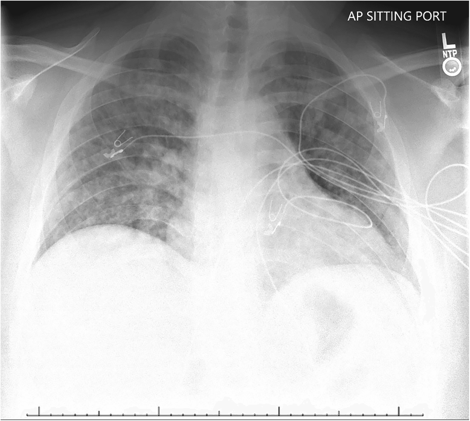
Figure 1. Chest X-ray image.
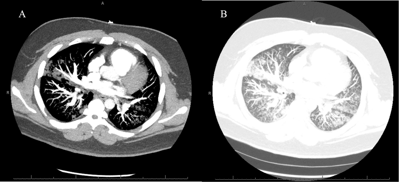
Figure 2. Computed tomography of the chest with contrast of axial view. (A) Soft tissue window and (B) lung window.
Toxicology was consulted because of the chlorine exposure. The toxicologist noted high suspicion for acute hypoxemic respiratory failure secondary to chlorine poisoning due to the timing of onset of symptoms, but mentioned that most patients experience complete resolution of pulmonary symptoms within 72 hours of exposure. Chest pain and substernal soreness were attributed to the possible presence of tracheobronchitis. Recommendation was made for supportive treatment involving bronchodilators, humidified oxygen, and a trial of steroids and possible 5% nebulized bicarbonate. Cardiology was consulted because of the pleuritic chest pain as well as elevated troponins. The cardiologist recommended an echocardiogram for evaluation of left ventricular function, with concern for etiologies such as myocarditis.
On hospital day 1, the patient’s respiratory status showed slight improvement, and he was weaned to 1–2 L/min of oxygen via NC. A transthoracic echocardiogram revealed a left ventricle of normal size and wall thickness but with mildly reduced systolic function. The left ventricular ejection fraction (LVEF) was estimated at 40%–45%, and right ventricular systolic function appeared grossly normal (Fig. 3). The working diagnosis was heart failure with reduced ejection fraction, possibly due to toxic cardiomyopathy or myocarditis. Cardiology recommended initiating metoprolol XR 25 mg and losartan 12.5 mg, both administered orally once daily. A consultation with an imaging cardiologist was also recommended to assess the need for a cardiac magnetic resonance imaging (MRI). C-reactive protein (CRP) and erythrocyte sedimentation rate (ESR) were recommended for evaluation.
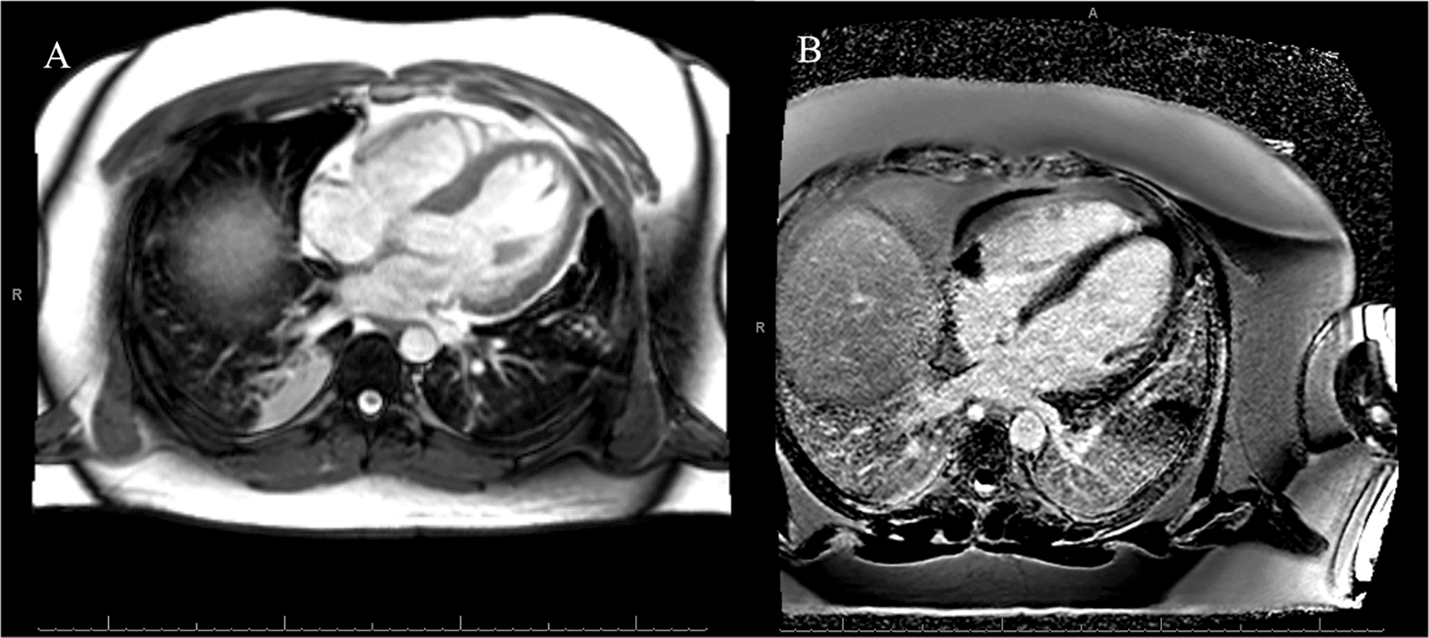
Figure 3. Cardiac magnetic resonance imaging findings in the myocardial wall. (A) T2-weighted and (B) T1-weighted post-contrast.
On hospital day 2, the patient remained stable on 1–2 L/min oxygen via NC. CRP and ESR levels were within normal limits. Cardiac MRI showed mildly to moderately reduced LVEF at 36% (≥47%) and reduced right ventricular ejection fraction at 35% (≥45%), with no evidence of myocarditis (Fig. 3). Cardiology advised continuing metoprolol and losartan, and adding spironolactone and empagliflozin.
By hospital day 3, the patient had been weaned off supplemental oxygen and was able to ambulate around the unit while maintaining an oxygen saturation level in the mid-1990s. He and his parents were counseled on the importance of weight loss for improving long-term respiratory function. A referral for nocturnal polysomnography was placed to evaluate for obstructive sleep apnea. The patient was discharged home the same day, with plans for outpatient follow-up with cardiology.
Discussion
This case highlights a rare instance of combined respiratory and cardiac dysfunction following acute exposure to Cl2 in a previously healthy young adult. While chlorine inhalation injury is well-documented for its pulmonary toxicity,13 the concomitant development of cardiac impairment remains a less recognized but clinically significant consequence. Cl2, a potent oxidizing agent, primarily affects mucosal surfaces and the respiratory tract, resulting in symptoms ranging from mild irritation to acute lung injury and acute respiratory distress syndrome.14 Most clinical manifestations occur within minutes of exposure and resolve within a few days with supportive therapy.3 Our patient exhibited typical respiratory symptoms, including cough, dyspnea, and hypoxemia, as well as radiographic findings consistent with chemical pneumonitis. His respiratory symptoms responded well to bronchodilators, corticosteroids, and supportive oxygen therapy, consistent with prior reports.
In contrast, cardiac involvement in cases like this is relatively uncommon in the literature. Studies have demonstrated that the adverse effects of inhaled toxic gases extend beyond the respiratory tract, potentially causing systemic endothelial dysfunction. This dysfunction may contribute to the development of cardiovascular conditions, including atherosclerosis and myocardial injury.15,16 Elevated troponin levels and reduced biventricular ejection fractions raised concern for chlorine-induced cardiotoxicity. While myocarditis was initially suspected, cardiac MRI showed no signs of inflammation. Takotsubo cardiomyopathy and viral myocarditis were considered but deemed unlikely based on imaging findings, absence of typical triggers, and lack of viral symptoms. These findings support a diagnosis of toxic cardiomyopathy.17 This case underscores the importance of early cardiology input and advanced imaging in assessing atypical cardiac symptoms after chemical exposure.
Management with standard heart failure medications, including beta-blockers, angiotensin receptor blockers, mineralocorticoid receptor antagonists, and sodium-glucose cotransporter-2 inhibitors, was initiated promptly and contributed to the patient’s clinical improvement.18 A limitation of this case is the absence of long-term follow-up data, which restricts our ability to assess the persistence or resolution of cardiac dysfunction over time.
Conclusion
This case highlights the potential for both respiratory and cardiac complications following acute exposure to Cl2. Prompt recognition and early supportive care, including heart failure management, were key to recovery. Clinicians should remain vigilant for cardiac involvement in similar toxic exposures.
Acknowledgments
We appreciate the patient’s cooperation in this study.
Funding Source
None.
Author Contributions
S.C. and H.C. contributed to drafting the manuscript. A.L. conducted the literature search, quality assessment, and data extraction. S.Y. was responsible for data revision. All authors have read and approved the final manuscript and agree with its content and data.
Data Availability
The datasets are available from the corresponding author upon reasonable request.
Ethical Statement
The patient gave written informed consent to the publication of this report and the accompanying images.
Conflict of Interest
The authors report no conflicts of interest in this work.
Supplemental Information
Supplemental information for this article can be found online at https://sup.jclinque.com/api/articles/75/download-suppl.
References
[1] Nambiema A, Coyo G, Barbe-Richaud JB, et al. Human chlorine gas exposition and its management—an umbrella review on human data. Crit Rev Toxicol. January 2022;52(1):32–50. doi:10.1080/10408444.2022.2035317.
[2] de Bruin-Hoegée M, van der Schans MJ, Langenberg JP, van Asten AC. Biomarker profiling in plants to distinguish between exposure to chlorine gas and bleach using LC-HRMS/MS and chemometrics. Forensic Sci Int. May 2024;358:112022. doi:10.1016/j.forsciint.2024.112022.
[3] Achanta S, Jordt SE. Toxic effects of chlorine gas and potential treatments: a literature review. Toxicol Mech Methods. May 2021;31(4):244–256. doi:10.1080/15376516.2019.1669244.
[4] Morim A, Guldner GT. Chlorine gas toxicity. In: StatPearls. Treasure Island (FL): StatPearls Publishing; June 26, 2023.
[5] White CW, Martin JG. Chlorine gas inhalation: human clinical evidence of toxicity and experience in animal models. Proc Am Thorac Soc. July 2010;7(4):257–263. doi:10.1513/pats.201001-008SM.
[6] Atalla A, Shulman J, Rose J, Lynch M. Trends in chlorine and chloramine gas exposures reported to United States poison centers. Clin Toxicol (Phila). September 2024;62(9):589–595. doi:10.1080/15563650.2024.2390139.
[7] Mashak KA, Agin K, Emaratkar E, Gholami Fesharaki M, Namdar H. The effect of wet cupping on reactive airway dysfunction syndrome (RADS) caused by early exposure to chlorine gas: a randomized clinical trial. Avicenna J Phytomed. January–February 2025;15(2):997–1015. doi:10.22038/ajp.2024.25107.
[8] Bleifuss W, Pepin L, Lock S, Kaluzniak H, Kempainen R. Chlorine gas induced acute respiratory distress syndrome due to pool shock. Am J Emerg Med. April 2, 2025;2(4):257. doi:10.1016/j.ajem.2025.04.001.
[9] Gülaşti F, Gülaşti S. Acute atrial fibrilation after inhalation of chlorine gas. GKD Anest Yoğ Bak Dern Derg. 2020;26(1):45–49. doi:10.5222/gkdad.2020.08941.
[10] Kose A, Kose B, Açikalin A, Gunay N, Yildirim C. Myocardial infarction, acute ischemic stroke, and hyperglycemia triggered by acute chlorine gas inhalation. Am J Emerg Med. October 2009;27(8):1022.e1–4. doi:10.1016/j.ajem.2008.12.029.
[11] Polat E, Demir MC. Myocardial Injury Due to Inhalation of A Mixture of Sodium Hypochlorite and Hydrochloric Acid. Sodyum Hipoklorit ve Hidroklorik Asit Karışımının İnhalasyonuna Bağlı Gelişen Miyokard Hasarı. Anatol J Emerg Med. December 2019;2(4):25–27.
[12] Rasouli HR, Hasani Nourian Y, Ghanei M, Salesi M, Nobakht Motlagh Ghoochani BF. A systematic review of the effect of acute, chronic, low, and high chlorine gas on people’s early and delayed complications. Iran Red Crescent Med J (IRCMJ). 2024;26(1). doi:10.22034/ircmj.2024.199016.
[13] Pham DT, Dao BN, Nguyen DT, et al. A rare case of acute respiratory distress syndrome due to chlorine gas inhalation: rapid progression with favorable ICU outcome. Respir Med Case Rep. 2025;53(3):102148. doi:10.1016/j.rmcr.2024.102148.
[14] Chen T-F, Wang C-H, Lain Hermes G, Ko W-J. Chlorine inhalation injury with acute respiratory distress syndrome treated by extra-corporeal membrane oxygenation system. Glob J Medical Clin Case Rep. 2020;4(11):005–009. doi:10.17352/2455-5282.000079.
[15] Zaky A, Ahmad A, Dell’Italia LJ, et al. Inhaled matters of the heart. Cardiovasc Regen Med. 2015;2. doi:10.14800/crm.997.
[16] Zaky A, Bradley WE, Lazrak A, et al. Chlorine inhalation-induced myocardial depression and failure. Physiol Rep. June 2015;3(6):e12439. doi:10.14814/phy2.12439.
[17] Amin HZ, Amin LZ, Pradipta A. Takotsubo cardiomyopathy: a brief review. J Med Life. January–March 2020;13(1):3–7. doi:10.25122/jml-2018-0067.
[18] Maddox TM, Januzzi JL Jr., Allen LA, et al. ACC expert consensus decision pathway for treatment of heart failure with reduced ejection fraction: a report of the american college of cardiology solution set oversight committee. J Am Coll Cardiol. April 2024;16(15):1444–1488. doi:10.1016/j.jacc.2023.12.024.
 |
Copyright: © 2025 Cho et al. This work is licensed under a Creative Commons Attribution 4.0 International License, which permits unrestricted use, distribution, and reproduction in any medium, provided the original work is properly cited. |