| Journal of Clinical Question. 2026; 3(1): e106 https://doi.org/10.69854/jcq.2025.0037 Advance access publication date 16 February 2026 |
 |
Review
Clonal Evolution in Myelodysplastic Syndromes: From Molecular Pathogenesis to Targeted Therapeutic Strategies
Nai-Bo Hu *
*
Hematology Department, Tianjin University Haihe Hospital, No. 890 Jinggu Road, Jinnan District, Tianjin 300350, China.
*Corresponding Author: e-mail: howareyoufeeling@163.com
Submitted: October 17, 2025 Accepted: February 15, 2026
Clinical Question Box
How does clonal evolution influence prognosis and therapeutic decision-making in patients with myelodysplastic syndromes?
Clonal evolution underlies the biological heterogeneity, disease progression, and treatment resistance observed in myelodysplastic syndromes. The accumulation and selection of genetic abnormalities shape clonal architecture, influencing prognosis and response to therapy. High-risk mutations and co-mutation patterns identify potential beneficiaries from early aggressive approaches, including targeted therapy or allogeneic stem cell transplantation. Molecular prognostic models such as the International Prognostic Scoring System-Revised to the Molecular International Prognostic Scoring System improve risk stratification by incorporating genetic data, but their static nature limits the prediction of disease trajectory, highlighting the importance of dynamic, longitudinal molecular monitoring to guide individualized treatment decisions.
Abstract
Myelodysplastic syndromes (MDS) constitute a heterogeneous group of clonal hematopoietic stem cell disorders characterized by ineffective hematopoiesis, peripheral cytopenias, and a significant risk of progression to acute myeloid leukemia. The core of MDS pathogenesis is a dynamic process of clonal evolution: initiating driver mutations in hematopoietic stem cells, under the combined pressure of genomic instability and the bone marrow microenvironment, leads to the continuous acquisition of new genetic abnormalities, forming a complex clonal architecture. This process is the fundamental cause of disease progression, therapeutic resistance, and clinical heterogeneity. This review systematically elaborates on the genetic and biological basis of clonal evolution, including frequent mutations and their interactions in key pathways such as epigenetic regulation, RNA splicing, and signal transduction. It analyzes how clonal competition, therapeutic selection pressure, and immune microenvironment editing drive clonal dynamics. Concurrently, the review details the evolution of prognostic assessment systems from the International Prognostic Scoring System-Revised to the Molecular International Prognostic Scoring System (IPSS-M), marking a revolutionary shift into the molecular era, and discusses both the clinical value and existing limitations of IPSS-M. Building upon this, the article comprehensively depicts the advancement of treatment strategies from immune modulation and epigenetic intervention to the era of precision targeting, integrating the latest evidence and optimized approaches for both lower- and higher-risk MDS. Finally, it proposes that future MDS management must integrate dynamic clonal monitoring, biomarker-based strategies, and multidisciplinary collaboration to achieve individualized and precise whole-course MDS management.
Keywords: Myelodysplastic syndromes, Clonal evolution, Gene mutation, IPSS-M, Hypomethylating agents, Targeted therapy
Introduction
The diagnostic framework and conceptual understanding of myelodysplastic syndromes (MDS) have evolved substantially. Once considered refractory anemia or a preleukemic condition, MDS is now recognized as a heterogeneous group of clonal hematopoietic neoplasms.1 Both the World Health Organization Classification of Tumours of Haematopoietic and Lymphoid Tissues, 5th Edition (WHO-HAEM5) and the International Consensus Classification (ICC) have adopted the term myelodysplastic neoplasms, underscoring their neoplastic nature.2,3 Unlike acute myeloid leukemia (AML), MDS originate not from a single fully transformed leukemic stem cell but from a genetically unstable hematopoietic stem and progenitor cell clone undergoing continuous evolution.4 The disease course is therefore defined by clonal evolution, characterized by the stepwise accumulation and competition of somatic mutations and cytogenetic abnormalities, resulting in complex oligoclonal architectures.5
Clonal evolution is central to the pathophysiology of MDS, driving disease heterogeneity, influencing progression risk, and shaping therapeutic response.6 Under treatment-induced selective pressures, dominant subclones may emerge through immune escape mechanisms or the acquisition of resistance-conferring mutations, ultimately leading to relapse or disease progression.7 Consequently, elucidating clonal evolutionary dynamics is essential for predicting disease trajectories, understanding mechanisms of treatment failure, and informing the development of novel therapeutic strategies. Advances in MDS therapy have paralleled increasing biological insight into disease pathogenesis, with prognostic assessment evolving from morphology-based classification systems toward integrated models incorporating cytogenetic and molecular features.8 In parallel, treatment approaches have progressed from predominantly supportive care and immunomodulatory therapies to epigenetic agents and targeted therapies directed against specific genetic alterations or signaling pathways.9 Despite these advances, allogeneic hematopoietic stem cell transplantation (allo-HSCT) remains the only potentially curative treatment, with clinical outcomes closely linked to the underlying clonal architecture and patterns of clonal evolution.10
MDS: Its Clonal Nature and Genetic Basis
Clonal Origin and Evolutionary Drivers
MDS are clonal disorders arising from hematopoietic stem and progenitor cells, characterized by pervasive genomic instability, including telomere dysfunction and impaired DNA damage–repair mechanisms.11 This instability provides the substrate for clonal evolution by promoting an increased mutational burden. Early in the disease course, clonal abnormalities may be limited to morphological dyshematopoiesis; however, with progression, particularly during transformation to higher-risk MDS or AML, the genetic features of dominant clones become increasingly apparent.5 Clonal evolution in MDS is driven by the interplay of intrinsic and extrinsic forces. Intrinsically, the continuous acquisition of genetic lesions fuels clonal diversification, whereas extrinsic selective pressures, most notably therapeutic interventions and the bone marrow microenvironment, shape clonal competition, fitness, and evolutionary trajectories.12
Genetic Landscape: Mutational Spectrum and Clinical Relevance
Somatic mutations are detected in over 90% of MDS patients, with more than half harboring multiple concurrent mutations, underscoring the marked genetic complexity of the disease.13 These alterations cluster within several key biological pathways (Fig. 1):
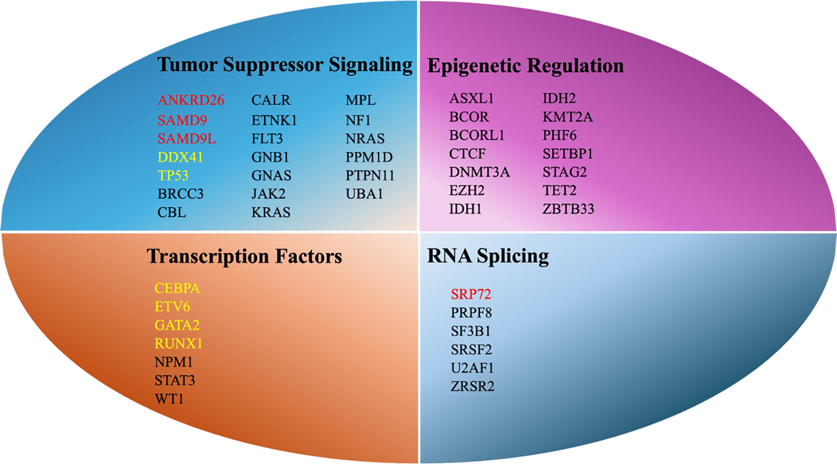
Figure 1. Key Mutational Pathways in MDS. In mutation annotation, black usually indicates somatic mutations, while red clearly denotes gemline mutations (requiring further confirmation with VAF); yellow may represent either somatic or germline mutations and requires comprehensive evaluation based on the specific locus and VAF. ASXL1, additional sex combs–like transcriptional regulator 1; DDX41, DEAD-box helicase 41; EZH2, enhancer of zeste homolog 2; IDH, isocitrate dehydrogenase; NRAS, NRAS proto-oncogene, GTPase; RUNX1, runt-related transcription factor 1; SAMD9/SAMD9L, sterile alpha motif domain–containing 9/sterile alpha motif domain–containing 9-like; SF3B1, splicing factor 3B subunit 1; SRSF2, serine/arginine-rich splicing factor 2; STAT3, signal transducer and activator of transcription 3; TET2, ten–eleven translocation methylcytosine dioxygenase 2; TP53, tumor protein p53; U2AF1, U2 small nuclear RNA auxiliary factor 1; ZRSR2, zinc finger RNA–binding motif and serine/arginine-rich protein 2; VAF, variant allele frequency; WT1, Wilms' tumor 1.
Ribonucleic Acid (RNA) Splicing Machinery
Mutations affecting components of the RNA splicing machinery, including splicing factor 3B subunit 1 (SF3B1), serine/arginine-rich splicing factor 2 (SRSF2), U2 small nuclear RNA auxiliary factor 1 (U2AF1), and zinc finger RNA–binding motif and serine/arginine-rich protein 2 (ZRSR2), are among the most frequent genetic alterations in MDS. Mutations in SF3B1 are closely associated with the presence of ring sideroblasts (RSs) and are generally linked to a favorable prognosis; however, this advantage is abrogated by specific co-occurring mutations.14 For instance, the co-mutation of SF3B1 with genes such as runt-related transcription factor 1 (RUNX1), BCL6 corepressor (BCOR), BCOR–like 1 (BCORL1), NRAS proto-oncogene, GTPase (NRAS), SRSF2, or stromal antigen 2 (STAG2) defines a molecular subset with significantly worse outcomes compared to SF3B1-mutant cases without these alterations.15 Similarly, emerging evidence suggests that deletion of the long arm of chromosome 5 (del(5q)), combined with an SF3B1 mutation, may also identify a subgroup with an independently poor prognosis, a risk interaction not yet fully captured by current prognostic models like the IPSS-M.16 Mutations in ZRSR2, which occur more frequently in male patients due to its X-linked location, are often associated with relatively favorable clinical outcomes, although their prognostic significance must be interpreted in the broader mutational context.17
Epigenetic Regulators
Epigenetic dysregulation represents a central pathogenic mechanism in MDS and involves mutations in genes regulating DNA methylation and chromatin structure.18 Key regulators of DNA methylation include ten–eleven translocation methylcytosine dioxygenase 2 (TET2) and DNA methyltransferase 3 alpha (DNMT3A), whereas epigenetic regulation of chromatin structure is mediated by histone-modifying genes such as additional sex combs–like transcriptional regulator 1 (ASXL1), enhancer of zeste homolog 2 (EZH2), and lysine methyltransferase 2A.19 Although mutations in DNMT3A, TET2, and ASXL1 may also be detected in age-related clonal hematopoiesis and, therefore, lack independent diagnostic specificity, they exert substantial biological effects in MDS. Particularly, mutations in DNMT3A and ASXL1 are well-established adverse prognostic markers associated with inferior survival.20
Transcription Factors
Mutations in hematopoietic transcription factors, including RUNX1, GATA binding protein 2 (GATA2), CCAAT/enhancer-binding protein alpha, and ETS variant transcription factor 6 (ETV6), disrupt normal hematopoietic differentiation and contribute to genomic instability.21 Notably, germline mutations in GATA2 and ETV6 underlie inherited MDS and AML predisposition syndromes, highlighting the role of transcriptional dysregulation in both sporadic and familial disease.22
Tumor Suppressor and Signaling Pathway Genes
Alterations in tumor suppressor genes and signaling pathways are strongly associated with disease aggressiveness. Mutations in tumor protein p53 (TP53) represent the most powerful adverse prognostic factor in MDS. Biallelic inactivation of TP53 is frequently associated with complex karyotypes, profound resistance to therapy, and extremely poor overall survival. Additionally, mutations affecting signaling pathways, including those involving rat sarcoma viral oncogene homolog (RAS) family members, confer proliferative and survival advantages to malignant clones.23
Importantly, the clinical impact of individual mutations is highly dependent on their combinatorial patterns. For instance, the favorable prognostic effect associated with SF3B1 mutations is lost when co-occurring with alterations in RUNX1, BCOR, BCORL1, neuroblastoma RAS viral oncogene homolog, SRSF2, or STAG2. Similarly, in MDS with deletion of the long arm of chromosome 5, coexisting TP53 mutations predict poor responses to lenalidomide and inferior clinical outcomes.24 The biological mechanisms underlying these adverse mutational interactions remain incompletely defined.
Germline Predisposition and Clonal Evolution
A subset of MDS arises from a defined germline predisposition and represents a biologically distinct entity. Germline mutations in GATA2 and sterile alpha motif domain–containing 9/sterile alpha motif domain–containing 9-like (SAMD9/SAMD9L) markedly increase the risk of MDS and AML, and are frequently associated with syndromic immunodeficiency. GATA2 deficiency and SAMD9/SAMD9L syndromes disrupt the development and function of myeloid and lymphoid immune cells, including monocytes, dendritic cells, and natural killer cells, resulting in profound impairment of both innate and adaptive immunity.25 Affected individuals are highly susceptible to refractory and recurrent infections, such as nontuberculous mycobacterial disease, invasive fungal infections, including aspergillosis, and severe viral infections including Epstein–Barr virus and human papillomavirus.26 These infections are a primary direct cause of mortality and, critically, promote the establishment of a chronically inflammatory bone marrow microenvironment. This sustained immunoinflammatory state functions as a powerful extrinsic driver that accelerates clonal evolution and perpetuates a self-reinforcing cycle linking immune deficiency, infection, inflammation, and clonal progression.27 These genes are essential for normal immune development, and their disruption results in profound innate and adaptive immune deficiency, predisposing affected individuals to recurrent mycobacterial, invasive fungal, and oncogenic viral infections.28 The resulting chronically inflammatory bone marrow microenvironment imposes strong selective pressure that accelerates clonal evolution.
In SAMD9/SAMD9L-associated disease, clonal evolution often involves somatic genetic rescue, including reversion mutations or uniparental disomy of chromosome 7q, which transiently alleviates the deleterious effects of the germline mutation.29 However, this unstable equilibrium reflects marked genomic instability and predisposes to subsequent acquisition of leukemogenic lesions, particularly monosomy 7 or deletion of chromosome 7q and activating RAS pathway mutations, ultimately accelerating disease progression.30
Leukemia-Defining Molecular Subtypes
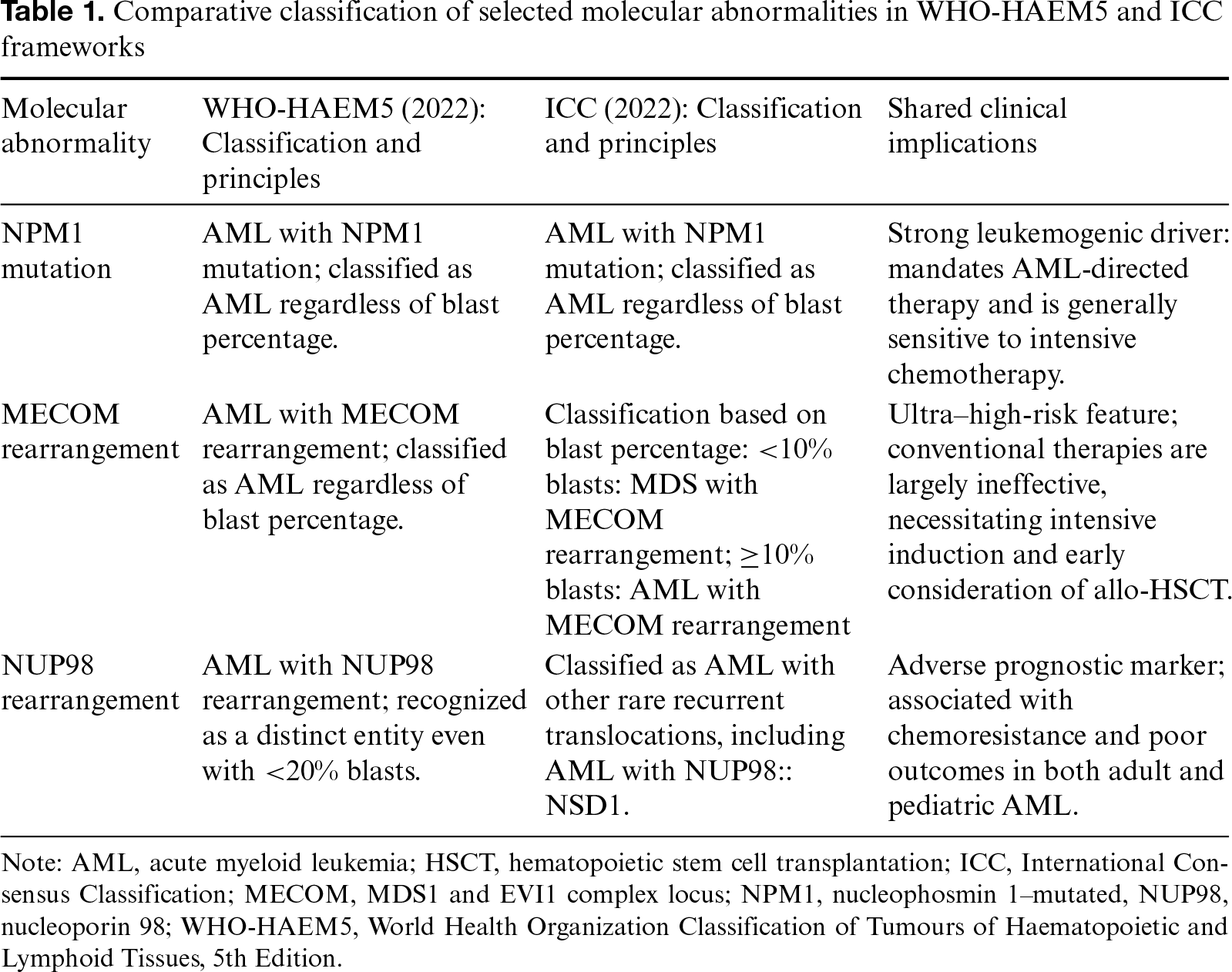
With advances in molecular understanding of MDS, contemporary classification systems have entered a genome-first era, in which certain molecular abnormalities supersede morphologic features, particularly blast percentage, as defining diagnostic criteria.2,3 Identification of these leukemia-defining molecular lesions is central to precision diagnosis and directly guides therapeutic strategy, most notably in the nucleophosmin 1 (NPM1)–mutated, MDS1 and EVI1 complex locus (MECOM)–rearranged, also called ecotropic viral integration site 1 (EVI1)–rearranged, and nucleoporin 98 (NUP98)–rearranged subtypes (Table 1).31
NPM1-Mutated Subtype
Both WHO-HAEM5 and ICC define AML by the presence of an NPM1 mutation, irrespective of bone marrow blast percentage (designated AML with mutated NPM1 in the ICC).2,3 This approach reflects strong evidence that NPM1 mutations drive a potent leukemogenic program, with clinical behavior and treatment responsiveness comparable to conventional blast-defined AML. Even when presenting with MDS-like morphology, patients typically respond favorably to standard intensive AML induction therapy, consisting of continuous-infusion cytarabine for 7 days combined with an anthracycline administered for 3 days.32 Such patients achieve high complete remission rates and derive clear benefit from subsequent consolidation chemotherapy and/or allo-HSCT. Overall outcomes closely mirror those of typical AML and are clearly distinct from other high-risk MDS subtypes.33
MECOM-Rearranged Subtype
WHO-HAEM5 recognizes AML with MECOM rearrangement as a distinct entity and mandates an AML diagnosis whenever a MECOM rearrangement is detected, regardless of blast percentage.2 In contrast, ICC applies blast-based stratification, classifying cases with <10% blasts as MDS with MECOM rearrangement and those with ≥10% blasts as AML with MECOM rearrangement.3 Despite these differences, both systems uniformly identify MECOM rearrangement as an ultra–high-risk feature, associated with poor therapeutic response and adverse outcomes.
NUP98-Rearranged Subtype
WHO-HAEM5 newly defines AML with NUP98 rearrangement as a distinct entity, allowing an AML diagnosis solely based on a NUP98 rearrangement, even when blasts are <20%.2 ICC classifies these cases under AML with other rare recurrent translocations.3 In East Asian adult AML cohorts, NUP98 rearrangements occur in approximately 5% of cases and are strongly associated with chemoresistance and poor clinical outcomes, underscoring their prognostic significance.34
Evolution of Prognostic Assessment Systems
The Transformative Significance of IPSS-M
Accurate prognostic stratification is central to individualized treatment in MDS. The IPSS and its revised version (IPSS-R) stratify risk based on bone marrow blast percentage, cytogenetics, and cytopenias, with IPSS-R remaining the most extensively validated model in clinical practice.35 The IPSS-M, introduced in 2022, builds on IPSS-R by incorporating the mutational status of 31 genes, thereby ushering MDS prognostication into the molecular era.24 Real-world studies show that IPSS-M reclassifies approximately 34% of patients, refining the heterogeneous IPSS-R intermediate-risk group into “moderate-low” and “moderate-high” categories and improving risk precision.36 Importantly, IPSS-M identifies occult high-risk disease among patients deemed low risk by IPSS-R due to adverse mutations such as biallelic TP53 inactivation or ASXL1 mutations, supporting earlier or more aggressive intervention.37 Emerging data further indicate that IPSS-M outperforms IPSS-R in predicting outcomes after allogeneic HSCT, enabling more accurate molecular guidance for transplant timing and candidacy.38
Clinical Limitations of IPSS-M
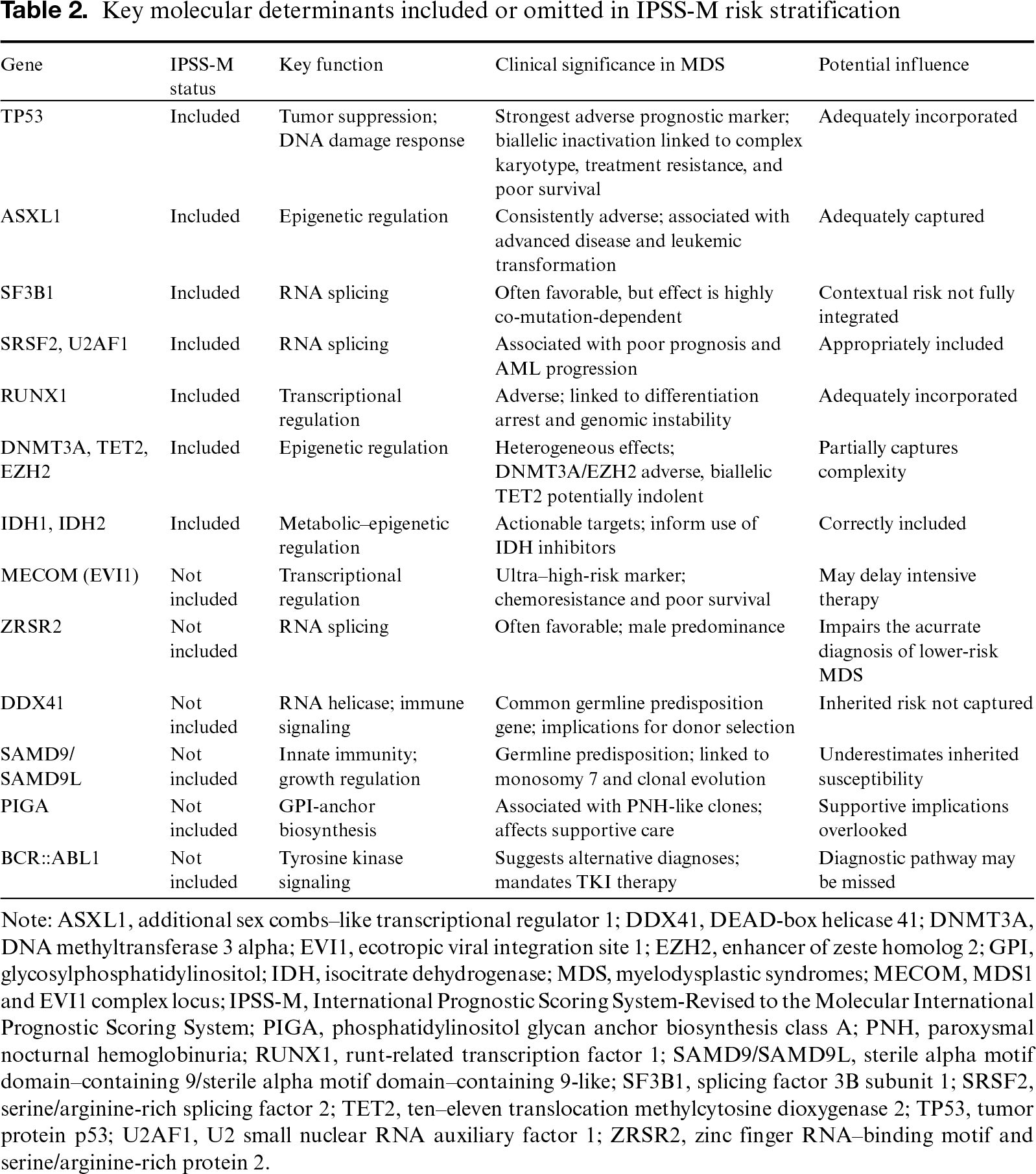
Despite its paradigm-shifting role in molecular risk stratification, the IPSS-M has several important limitations in clinical practice. Although it incorporates the mutational status of 31 genes, gene coverage remains incomplete. Several clinically relevant genes with established prognostic or therapeutic implications in MDS are excluded, including MECOM, ZRSR2, Fanconi anemia complementation group L, and phosphatidylinositol glycan anchor biosynthesis class A (PIGA) (Table 2). Additionally, key germline predisposition genes such as DEAD-box helicase 41 (DDX41) and SAMD9/SAMD9L are not included, potentially leading to underestimation of risk and omission of inherited susceptibility. Such oversights may cause clinical misjudgment and a delay in appropriate therapy. For instance, conventional karyotyping is insufficient to detect MECOM/EVI1 rearrangements due to limitations in resolution and metaphase quality. Thus, the omission of this ultra–high-risk feature may lead to significant underestimation of risk and delay intensive therapy like transplantation.39
A second major limitation is the insufficient modeling of gene–gene interactions and clonal dynamics. IPSS-M assigns weighted risk scores to individual mutations but does not fully capture context-dependent effects of co-mutation patterns, such as the variable prognostic impact of SF3B1 mutations in specific genetic backgrounds.40 More fundamentally, IPSS-M represents a static molecular snapshot at diagnosis, whereas MDS is characterized by continuous clonal evolution driven by genomic instability and treatment-related selective pressures, limiting accurate prediction of disease trajectory.41
Relatedly, IPSS-M does not comprehensively integrate mutational burden or variant allele frequency dynamics, which provide critical insights into clonal size, treatment response, and emerging resistance. Although the allelic state is partially considered for selected genes such as TP53, this level of granularity is not uniformly applied across other prognostically relevant mutations, including STAG2 and RUNX1. Further, IPSS-M does not consider clonal architecture, a defining feature of MDS reflecting hierarchical organization and dynamic subclonal evolution during therapy.41
Finally, practical and conceptual limitations affect implementation. IPSS-M requires comprehensive next-generation sequencing, increasing technical complexity, cost, and turnaround time compared to IPSS-R.42 Moreover, it does not incorporate other prognostically relevant factors such as bone marrow fibrosis, immune dysfunction, inflammatory microenvironmental features, age, or comorbidities, all of which influence treatment tolerance and real-world clinical decision-making.
Optimizing Treatment Strategies for Lower-Risk MDS
In patients with lower-risk MDS, anemia management remains the central therapeutic priority. Effective correction of anemia is critical not only for improving quality of life and reducing transfusion dependence but also for conferring long-term survival benefits.43 Historically, treatment strategies relied predominantly on supportive care measures and erythropoiesis-stimulating agents (ESAs). However, advances in the understanding of erythroid dysregulation have led to the development of novel agents that more directly target ineffective erythropoiesis and disease biology.
Erythroid Maturation Agents
Luspatercept, a transforming growth factor-β (TGF-β) superfamily ligand trap, enhances late-stage erythroid maturation by inhibiting aberrant SMAD2/3 signaling. Its clinical efficacy was established in the phase III MEDALIST trial, which demonstrated significant benefit in ESA–refractory, transfusion-dependent lower-risk MDS with RSs.44 Subsequently, the phase III COMMANDS trial showed superior efficacy of luspatercept compared to ESAs in treatment-naïve, transfusion-dependent lower-risk MDS, supporting its use as a first-line therapeutic option.45 Long-term follow-up from MEDALIST confirmed improved overall survival among patients achieving transfusion independence for at least 8 weeks, and real-world data published in 2025 reported a 75% reduction in mortality risk among hematologic responders compared to non-responders.46,47
KER-050 (elritercept), a next-generation activin receptor type IIA ligand trap, extends this therapeutic approach by inhibiting activin A and related TGF-β superfamily ligands.48 Early-phase clinical studies have demonstrated encouraging efficacy and favorable safety profiles in patients with lower- to intermediate-risk MDS, positioning KER-050 as a promising addition to anemia-directed treatment strategies.49
Disease-Modifying Approaches
Imetelstat, a first-in-class telomerase inhibitor, has emerged as a potential disease-modifying therapy in lower-risk MDS. In the phase III IMerge trial, imetelstat significantly increased rates of transfusion independence in ESA-refractory patients. Importantly, treatment was associated with reductions in AML transformation risk and declining variant allele frequencies of driver mutations, suggesting biological activity beyond symptomatic anemia control.50
Iron Chelation and Supportive Care
Besides pharmacological therapies, optimal supportive care remains a critical component of management. Treatment of transfusion-related secondary iron overload with iron chelation therapy has been consistently associated with improved survival in lower-risk MDS.51 Collectively, these data underscore that effective anemia control, through erythroid maturation agents, emerging disease-modifying therapies, and proactive management of iron overload, remains a cornerstone for optimizing long-term outcomes in lower-risk MDS.
Exploration of Spliceosome Modulation
Direct targeting of the spliceosome, a core machinery frequently mutated in MDS, has been explored. H3B-8800, an oral small molecule modulator of the SF3b complex, demonstrated the potential to induce transfusion independence in a subset of patients with spliceosome-mutant, lower-risk MDS in a phase I trial.52 However, overall efficacy was modest, with no standard complete or partial remissions observed, and clinical development has been discontinued.53 This experience validates the spliceosome as a therapeutic target but highlights the challenges in achieving robust clinical efficacy with single-agent modulation.
In the management of lower-risk MDS, adequate doses of ESAs remain the classic first-line therapy for patients with serum erythropoietin (EPO) levels <500 IU/L.54 Luspatercept has also emerged as an established first-line option, particularly for patients with SF3B1 mutations or RS-associated MDS, in whom it demonstrates favorable efficacy in those with EPO levels between 200 and 500 IU/L and even <200 IU/L.55 In addition, the MEDALIST trial showed activity in non-RS, non-SF3B1 lower-risk MDS.44 The phase 3 COMMANDS trial further demonstrated superior erythroid responses with luspatercept compared with darbepoetin alfa in ESA-naive, transfusion-dependent lower-risk MDS, establishing luspatercept as a frontline option for a broader patient population irrespective of RS status.45 In real-world practice, the choice between luspatercept and ESAs should be individualized according to local reimbursement policies and patient financial considerations.
Imetelstat is indicated for transfusion-dependent lower-risk MDS patients who have failed, lost response to, or are ineligible for ESAs.50 It is particularly valuable in patients with baseline EPO >500 IU/L, a population with an anticipated poor response to ESAs and reduced benefit from luspatercept. The US Food and Drug Administration approval was based on the IMerge phase 3 trial, and imetelstat is now included as a National Comprehensive Cancer Network Category 1 preferred option for this indication.56
For patients with del(5q) MDS, lenalidomide remains the standard of care.57 However, its efficacy is attenuated with increasing TP53 mutation burden. In del(5q) patients harboring TP53 mutations, a sequential strategy using frontline hypomethylating agents (HMAs) to reduce the TP53 clonal burden—followed by lenalidomide—has shown potential efficacy in a recently reported case, suggesting a novel therapeutic approach for this challenging subset.58
For patients with EPO >500 IU/L who exhibit clinical features predictive of response to immunosuppressive therapy, including age ≤60 years, bone marrow blasts ≤5%, hypocellular marrow, presence of a paroxysmal nocturnal hemoglobinuria clone, or signal transducer and activator of transcription 3–mutant cytotoxic T-cell clones, immunosuppressive therapy combined with thrombopoietin receptor agonists may achieve favorable outcomes.59 When these strategies are ineffective, treatment with hypomethylating agents or enrollment in clinical trials should be considered. For patients with significantly compromised quality of life, early evaluation for HSCT is recommended.60
Optimizing Treatment Strategies for Higher-Risk MDS
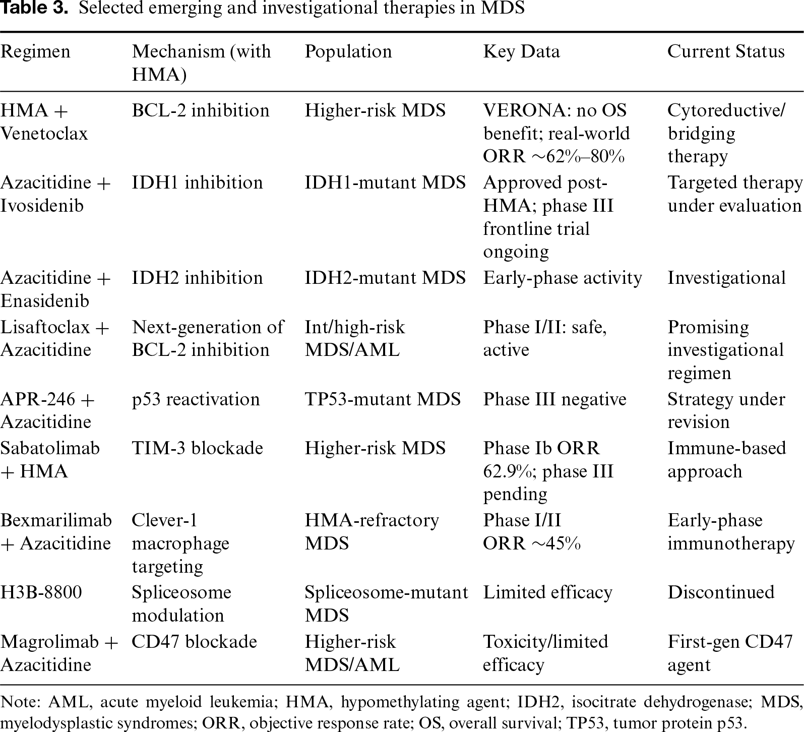
The primary therapeutic goals in higher-risk MDS (encompassing moderate–high-, high-, and very–high-risk categories) are to delay disease progression, eradicate malignant clones, and prevent transformation to AML. While HMAs such as azacitidine and decitabine (including the oral fixed-dose combination of decitabine with cedazuridine) have long served as the therapeutic backbone, treatment paradigms are rapidly evolving toward mechanism-driven combination regimens and precision-based strategies designed to overcome resistance and address high-risk disease biology (Table 3).
Refinement and Expansion of Combination Therapies
HMA Plus BCL-2 Inhibition
Combination therapy with azacitidine or decitabine plus the BCL-2 inhibitor venetoclax, adapted from its success in AML, has demonstrated rapid and deep cytoreductive responses in high-risk MDS. Across real-world and early clinical studies, overall response rates of 62%–80% have been reported, and this regimen is widely used as an intensified strategy or bridge to allo-HSCT.61,62 However, in the randomized phase III VERONA trial, the addition of venetoclax to HMA therapy failed to confer a statistically significant improvement in overall survival compared to HMA monotherapy (hazard ratio, 0.908).63 Although higher response rates were observed in molecularly adverse subsets such as TP53-mutated disease, survival benefits remained modest. Consequently, HMA–venetoclax is best regarded as an effective disease-modifying and bridging regimen rather than a new standard first-line therapy.
HMA Plus Isocitrate Dehydrogenase (IDH) Inhibitors
Targeting mutant IDH constitutes a precision-medicine strategy applicable to approximately 3%–13% of patients with MDS.64,65 In IDH1-mutant disease, the IDH1 inhibitor ivosidenib is approved for relapsed or refractory MDS following failure of HMA therapy.66 Its combination with azacitidine is currently being evaluated in the frontline setting in the randomized phase III PyramIDH trial.67.
Similarly, the IDH2 inhibitor enasidenib is under investigation as monotherapy and in combination with hypomethylating agents across multiple disease settings.68,69 Early-phase clinical studies have demonstrated feasibility and biological activity, although definitive efficacy data and survival outcomes require further validation in larger controlled trials.70. These approaches exemplify the ongoing shift toward genomically informed treatment selection in higher-risk MDS.
Next-Generation BCL-2 Inhibitors
To address the resistance and toxicity associated with first-generation BCL-2 inhibition, next-generation BCL-2 inhibitors such as lisaftoclax are being developed. Lisaftoclax exhibits optimized selectivity and tolerability, and early phase I/II studies combining lisaftoclax with azacitidine in treatment–naïve higher-risk MDS and AML have demonstrated manageable safety profiles and encouraging antileukemic activity.71 This approach represents a promising evolution of BCL-2–based therapy.
TP53-Directed Therapy
TP53-mutated MDS, frequently associated with complex karyotypes, represent one of the most adverse prognostic subtypes, largely resistant to conventional therapies and constituting a major unmet clinical need.72
Efforts to directly restore p53 function have thus far produced mixed results. Notably, the phase III trial evaluating the p53 reactivator APR-246 (eprenetapopt) in combination with azacitidine failed to achieve its primary endpoints of complete remission and overall survival improvement.73 Consequently, therapeutic strategies are increasingly shifting toward intensified combination approaches, including triplet regimens incorporating APR-246, azacitidine, and venetoclax. In contrast, MDM2 inhibitors, which are restricted to TP53–wild-type disease, have demonstrated limited clinical activity in MDS, prompting growing interest in next-generation approaches such as PROTAC-based targeted protein degraders.74
Modulation of the Immune Microenvironment
Targeting the immunosuppressive bone marrow microenvironment has emerged as an important complementary therapeutic strategy in higher-risk MDS. Immune checkpoint modulation through TIM-3 inhibition has revealed encouraging early activity; notably, the anti–TIM-3 antibody sabatolimab, when combined with HMAs, demonstrated favorable tolerability and an overall response rate of 62.9% in a phase Ib study, and the randomized phase III STIMULUS-MDS2 trial has completed accrual with results awaited.75,76 In parallel, macrophage-directed approaches are gaining traction, exemplified by bexmarilimab, an antibody targeting Clever-1 on immunosuppressive macrophages, which, in combination with azacitidine, achieved a preliminary overall response rate of 45% in HMA–refractory higher-risk MDS, highlighting the therapeutic potential of macrophage reprogramming within the bone marrow niche.77 However, first-generation CD47 blockade strategies (e.g., magrolimab in combination with HMAs) have been constrained by dose-limiting toxicities and inconsistent clinical efficacy.78
Allo-HSCT
Allo-HSCT remains the only potentially curative therapy for MDS.79 Long-term outcomes indicate a 5-year overall survival of approximately 33.9%–52.8% following transplantation.80. Optimal transplant outcomes depend on careful patient selection, effective pre-transplant disease debulking, and posttransplant surveillance, including minimal residual disease (MRD) monitoring using high-throughput sequencing to enable timely preemptive intervention.81
Risk-Based Transplant Selection
The IPSS-M scoring system has improved identification of patients most likely to benefit from allo-HSCT; however, accurate diagnostic classification remains the cornerstone of transplant decision-making. Molecular and cytogenetic features increasingly guide both the indication and timing of transplantation, emphasizing the need for integrated clinicogenomic assessment.24 Under the WHO-HAEM5 classification, cases harboring NPM1 mutations, MECOM rearrangements, or NUP98 rearrangements are now classified as AML or distinct AML subtypes rather than MDS. For these patients, frontline management should follow AML treatment principles, including intensive induction chemotherapy and early incorporation of allo-HSCT, with urgency comparable to that applied in high-risk AML.
In patients meeting diagnostic criteria for MDS, decisions regarding allo-HSCT should be individualized according to molecular subtype. Indolent subtypes, including isolated SF3B1 mutation and biallelic TET2 mutations, generally do not warrant allo-HSCT.82 Subtypes requiring individualized consideration, such as del(5q) with high-risk co-mutations, DDX41-mutated disease, and U2AF1 or SRSF2 mutations, require integration of molecular features, blast burden, and clinical factors to guide transplant timing.83 Conversely, high-risk molecular subtypes, including TP53-mutated disease with complex karyotype, −7/del(7q) with SETBP1 or RAS mutations, and EZH2–ASXL1 co-mutations, strongly favor early allo-HSCT and consideration of clinical trial enrollment when feasible.84
Germline predisposition syndromes substantially complicate allo-HSCT decision-making. Patients with germline susceptibility (e.g., GATA2 or SAMD9/SAMD9L mutations) frequently exhibit baseline immune dysfunction, recurrent infections, and organ impairment, necessitating highly individualized risk–benefit assessment.85 Donor selection is particularly challenging, as germline genetic screening is mandatory to avoid related donors carrying the same pathogenic variants.
Infection Control
Pre-transplant infection control represents a major challenge in patients with inherited predisposition syndromes. Active or refractory infections, particularly nontuberculous mycobacterial infections and invasive fungal diseases, may constitute contraindications to transplantation or carry an extremely high risk of transplant-related mortality.86 Achieving adequate infection control often requires prolonged, multidisciplinary antimicrobial therapy, which can delay allo-HSCT and increase the risk of disease progression; nevertheless, optimization of infectious status before conditioning remains essential, albeit frequently difficult.87
Posttransplant outcomes are further influenced by delayed immune reconstitution due to underlying immunodeficiency, resulting in high rates of severe infections, including bacteremia (22.3%) and pulmonary fungal infections (12.4%).88 Reduced tolerance to conditioning regimens, related to poor baseline performance status and prior infections, necessitates individualized conditioning intensity and tailored graft-vs.-host disease prophylaxis to balance disease control with transplant-related toxicity.
Further Direction
Future therapeutic strategies for MDS must be dynamic, precision-driven, and grounded in a comprehensive understanding of clonal evolution. Longitudinal monitoring of variant allele frequencies and clonal architecture using high-throughput sequencing enables response-adaptive treatment strategies, with post-treatment MRD status emerging as a powerful predictor of relapse and a critical guide for timely preemptive intervention.89 Moving beyond static risk models, integration of artificial intelligence–driven, multiparametric frameworks that incorporate clinical variables, molecular profiles, and imaging data holds promise for predicting clonal evolutionary trajectories and optimizing treatment sequencing, thereby enabling individualized, dynamic prognostic systems that surpass the limitations of current tools such as IPSS-M.90
Ultimately, effective control of clonal evolution will require rationally designed therapeutic combinations, administered concurrently or sequentially, to target both dominant and emerging subclones. Future drug development should prioritize novel and historically challenging targets, including transcription factors, downstream effectors of spliceosome dysregulation, and components of the bone marrow microenvironment, as well as synthetic lethality–based strategies to overcome adaptive resistance mechanisms.91
Conclusion
MDS represent a biologically heterogeneous group of disorders in which clinical outcomes are increasingly shaped by underlying molecular architecture and clonal dynamics. Though advances in genomic profiling, risk stratification, and targeted therapeutics have refined diagnosis and treatment selection, durable disease control remains limited for many high-risk subgroups. Future progress will depend on integrating precision medicine, dynamic MRD-guided strategies, and rational combination therapies to overcome clonal evolution and resistance, with allogeneic transplantation and clinical trial enrollment remaining essential components of curative and disease-modifying approaches.
Acknowledgment
None.
Funding Statement
None.
Ethics Approval Statement
Not applicable.
Patient Consent Statement
Not applicable.
Conflict of Interest Disclosure
The authors declare that they have no competing interests.
Data Sharing Statement
The raw data are available upon reasonable request to the corresponding author via email.
Generative AI Declaration
During the preparation of this manuscript, the authors used ChatGPT to assist with proofreading. All content was subsequently reviewed and edited by the authors, who assume full responsibility for the accuracy and integrity of the published work.
Authors’ Contributions
Nai-Bo Hu was responsible for study conceptualization, investigation, preparation of the original draft, project administration, and approved the final manuscript.
Supplemental Information
Supplemental information for this article can be found online at https://sup.jclinque.com/api/articles/106/download-suppl.
References
[1] Lapadat ME, Stanca O, Berbec NM, Negotei C, Colita A. Navigating the new era in myelodysplastic neoplasms: a review of prognostic implications of the IPSS-M score and 2022 WHO classification. Hematol Rep. October 30, 2025;17(6):58. doi:10.3390/hematolrep17060058.
[2] Loghavi S, Kanagal-Shamanna R, Khoury JD, et al. Fifth edition of the world health classification of tumors of the hematopoietic and lymphoid tissue: myeloid neoplasms. Mod Pathol. February 2024;37(2):100397. doi:10.1016/j.modpat.2023.100397.
[3] Arber DA, Orazi A, Hasserjian RP, et al. International consensus classification of myeloid neoplasms and acute leukemias: integrating morphologic, clinical, and genomic data. Blood. September 15, 2022;140(11):1200–1228. doi:10.1182/blood.2022015850.
[4] Cazzola M. Myelodysplastic syndromes. N Engl J Med. October 1, 2020;383(14):1358–1374. doi:10.1056/NEJMra1904794.
[5] García R, Alkayyali T, Gomez LM, et al. Recurrent cytogenetic abnormalities reveal alterations that promote progression and transformation in myelodysplastic syndrome. Cancer Genet. November 01, 2024;288–289:92–105. doi:10.1016/j.cancergen.2024.10.002.
[6] Schwind S, Jentzsch M, Kubasch AS, Metzeler KH, Platzbecker U. Myelodysplastic syndromes: biological and therapeutic consequences of the evolving molecular aberrations landscape. Neoplasia. November 01, 2021;23(11):1101–1109. doi:10.1016/j.neo.2021.09.002.
[7] Bernard E, Tuechler H, Greenberg PL, et al. Molecular international prognostic scoring system for myelodysplastic syndromes. NEJM Evid. July 2022;1(7):EVIDoa2200008. doi:10.1056/EVIDoa2200008.
[8] Chen X, Patkar N, Tembhare P, et al. Fifth edition WHO classification: myeloid neoplasms. J Clin Pathol. April 17, 2025;75(5):335–345. doi:10.1136/jcp-2024-210022.
[9] Karel D, Valburg C, Woddor N, Nava VE, Aggarwal A. Myelodysplastic neoplasms (MDSthe current and future treatment landscape. Current Oncol. 2024;31(4):1971–1993. doi:10.3390/curroncol31040148.
[10] Berg T, Salter B, Radford M, Chen HT, Leber B. Allogeneic stem cell transplantation: the relevance of conditioning regime intensity for myelodysplastic syndromes (MDS). Current Oncol. 2025;32(6):319. doi:10.3390/curroncol32060319.
[11] Rodriguez-Sevilla JJ, Adema V, Garcia-Manero G, Colla S. Emerging treatments for myelodysplastic syndromes: Biological rationales and clinical translation. Cell Rep Med. 2023;4(2):100940. doi:10.1016/j.xcrm.2023.100940.
[12] Villaume MT, Savona MR. Pathogenesis and inflammaging in myelodysplastic syndrome. Haematologica. February 1, 2025;110(2):283–299. doi:10.3324/haematol.2023.284944.
[13] Garcia-Manero G. Myelodysplastic syndromes: 2023 update on diagnosis, risk-stratification, and management. Am J Hematol. August 2023;98(8):1307–1325. doi:10.1002/ajh.26984.
[14] Huber S, Haferlach T, Meggendorfer M, et al. Mutations in spliceosome genes in myelodysplastic neoplasms and their association to ring sideroblasts. Leukemia. February 01, 2023;37(2):500–502. doi:10.1038/s41375-022-01783-y.
[15] Zemanova Z, Aghova T, Lhotska H, et al. SF3B1 gene mutations and their significance for patients with myelodysplastic neoplasms (MDS). Blood. November 05, 2024;144:6736. doi:10.1182/blood-2024-202114.
[16] Karel D, Valburg C, Woddor N, Nava VE, Aggarwal A. Myelodysplastic neoplasms (MDSthe current and future treatment landscape. Curr Oncol. April 3, 2024;31(4):1971–1993. doi:10.3390/curroncol31040148.
[17] Yacout M, Katamesh B, Jabban Y, et al. Characterisation and prognostic impact Of ZRSR2 mutations in myeloid neoplasms. Leukemia. December 01, 2024;38(12):2727–2730. doi:10.1038/s41375-024-02374-9.
[18] Bond DR, Lee HJ, Enjeti AK. Unravelling the epigenome of myelodysplastic syndrome: diagnosis, prognosis, and response to therapy. Cancers. 2020;12(11):3128. doi:10.3390/cancers12113128.
[19] Basquiera AL, Andreoli V, Grille S, Belli CB. Progress in the genetics of myelodysplastic syndromes with a latin American perspective. Genes. 2025;16(6):687. doi:10.3390/genes16060687.
[20] Yang L, Wei X, Gong Y. Prognosis and risk factors for ASXL1 mutations in patients with newly diagnosed acute myeloid leukemia and myelodysplastic syndrome. Cancer Med. January 01, 2024;13(1):e6871. doi:10.1002/cam4.6871.
[21] Kontandreopoulou C-N, Kalopisis K, Viniou N-A, Diamantopoulos P. The genetics of myelodysplastic syndromes and the opportunities for tailored treatments. Review. Front Oncol. October 20, 2022;12:1100. doi:10.3389/fonc.2022.989483.
[22] Abou Dalle I, Bannon SA, Patel KP, et al. Germline genetic predisposition to myeloid neoplasia from GATA2 gene mutations: lessons learned from two cases. JCO Precis Oncol. 2019;3:1–5. doi:10.1200/PO.18.00301.
[23] Huang N, Chang C, Wu L, et al. Characteristics and prognostic implications of TP53 mutations in Chinese patients with myelodysplastic syndromes. Br J Haematol. October 2025;207(4):1397–1407. doi:10.1111/bjh.70078.
[24] Bernard E, Tuechler H, Greenberg Peter L, et al. Molecular International prognostic scoring system for myelodysplastic syndromes. NEJM Evidence. June 28, 2022;1(7):EVIDoa2200008. doi:10.1056/EVIDoa2200008.
[25] Fabozzi F, Mastronuzzi A, Ceglie G, Masetti R, Leardini D. GATA 2 deficiency: focus on immune system impairment. Mini review. Front Immunol. June 13, 2022;13:865773. doi:10.3389/fimmu.2022.865773.
[26] Dancy E, Stratton P, Pichard DC, et al. Human papillomavirus disease in GATA2 deficiency: a genetic predisposition to HPV-associated female anogenital malignancy. Original Research. Front Immunol. August 29, 2024;15:1445711. doi:10.3389/fimmu.2024.1445711.
[27] Sahoo SS, Kozyra EJ, Wlodarski MW. Germline predisposition in myeloid neoplasms: unique genetic and clinical features of GATA2 deficiency and SAMD9/SAMD9L syndromes. Best Pract Res Clin Haematol. September 2020;33(3):101197. doi:10.1016/j.beha.2020.101197.
[28] Sahoo SS, Erlacher M, Wlodarski MW. Genetic and clinical spectrum of SAMD9 and SAMD9L syndromes: from variant interpretation to patient management. Blood. January 30, 2025;145(5):475–485. doi:10.1182/blood.2022017717.
[29] Sahoo SS, Pastor VB, Goodings C, et al. Clinical evolution, genetic landscape and trajectories of clonal hematopoiesis in SAMD9/SAMD9L syndromes. Nat Med. October 2021;27(10):1806–1817. doi:10.1038/s41591-021-01511-6.
[30] Erlacher M, Andresen F, Sukova M, et al. Spontaneous remission and loss of monosomy 7: a window of opportunity for young children with SAMD9L syndrome. Haematologica. February 1, 2024;109(2):422–430. doi:10.3324/haematol.2023.283591.
[31] Ally F, Chen X. Acute myeloid leukemia: diagnosis and evaluation by flow cytometry. Cancers. 2024;16(22):3855. doi:10.3390/cancers16223855.
[32] Itzykson R. NPM1-mutated AML: how many diseases? Blood. August 15, 2024;144(7):681–683. doi:10.1182/blood.2024025032.
[33] Chan O, Al Ali N, Tashkandi H, et al. Mutations highly specific for secondary AML are associated with poor outcomes in ELN favorable risk NPM1-mutated AML. Blood Adv. March 12, 2024;8(5):1075–1083. doi:10.1182/bloodadvances.2023011173.
[34] Kim N, Choi YJ, Cho H, et al. NUP98 is rearranged in 5.0% of adult East Asian patients with AML. Blood Adv. October 8, 2024;8(19):5122–5125. doi:10.1182/bloodadvances.2024012960.
[35] Greenberg PL, Tuechler H, Schanz J, et al. Revised international prognostic scoring system for myelodysplastic syndromes. Blood. September 20, 2012;120(12):2454–2465. doi:10.1182/blood-2012-03-420489.
[36] Sauta E, Robin M, Bersanelli M, et al. Real-world validation of molecular international prognostic scoring system for myelodysplastic syndromes. J Clin Oncol. May 20, 2023;41(15):2827–2842. doi:10.1200/jco.22.01784.
[37] Kasprzak A, Nachtkamp K, Gattermann N, Germing U. Assessing the prognosis of patients with myelodysplastic syndromes (MDS). Cancers (Basel). April 12, 2022;14(8):1941. doi:10.3390/cancers14081941.
[38] Chen H, Jiang S, Wei R, et al. Threefold IPSS-M reclassification outperforms original stratification in predicting post-transplant outcomes for MDS patients. Cell Transplant. January–December 2025;34:9636897251348406. doi:10.1177/09636897251348406.
[39] Polprasert C, Chanswangphuwana C, Owattanapanich W, et al. Clinical characteristics and outcomes of myeloid neoplasms with mecom rearrangement: results from a nationwide multicenter study. Blood. 2023;142(Supplement 1):4213. doi:10.1182/blood-2023-180100.
[40] Sauta E, Robin M, Bersanelli M, et al. Real-world validation of molecular international prognostic scoring system for myelodysplastic syndromes. J Clinic Oncol. May 20, 2023;41(15):2827–2842. doi:10.1200/JCO.22.01784.
[41] El Hussein S, Loghavi S. The impact of clonal hierarchy and heterogeneity on phenotypic manifestations of myelodysplastic neoplasms. Cancers. 2022;14(22):5690. doi:10.3390/cancers14225690.
[42] Lee W-H, Tsai M-T, Tsai C-H, et al. Validation of the molecular international prognostic scoring system in patients with myelodysplastic syndromes defined by international consensus classification. Blood Cancer J. August 09, 2023;13(1):120. doi:10.1038/s41408-023-00894-8.
[43] Meunier M, Park S. Lower-risk myelodysplastic syndromes: current treatment options for anemia. EJHaem. November 2022;3(4):1091–1099. doi:10.1002/jha2.523.
[44] Fenaux P, Platzbecker U, Mufti Ghulam J, et al. Luspatercept in patients with lower-risk myelodysplastic syndromes. New England J Med. January 09, 2020;382(2):140–151. doi:10.1056/NEJMoa1908892.
[45] Della Porta MG, Garcia-Manero G, Santini V, et al. Luspatercept versus epoetin alfa in erythropoiesis-stimulating agent-naive, transfusion-dependent, lower-risk myelodysplastic syndromes (COMMANDSprimary analysis of a phase 3, open-label, randomised, controlled trial. Lancet Haemat. September 01, 2024;11(9):e646–e658. doi:10.1016/S2352-3026(24)00203-5.
[46] Platzbecker U, Santini V, Komrokji RS, et al. Long-term utilization and benefit of luspatercept in transfusion-dependent, erythropoiesis-stimulating agent-refractory or -intolerant patients with lower-risk myelodysplastic syndromes with ring sideroblasts. Leukemia. November 01, 2023;37(11):2314–2318. doi:10.1038/s41375-023-02031-7.
[47] Consagra A, Lanino L, Al Ali NH, et al. Response to luspatercept can be predicted and improves overall survival in the real-life treatment of LR-MDS. Hemasphere. February 2025;9(2):e70086. doi:10.1002/hem3.70086.
[48] Chifotides HT, Duminuco A, Torre E, et al. Emerging therapeutic approaches for anemia in myelofibrosis. Curr Hematol Malig Rep. May 3, 2025;20(1):7. doi:10.1007/s11899-025-00751-4.
[49] Lachey J, Rovaldi C, Bobba S, et al. Elritercept, a modified activin receptor IIA ligand trap, increased erythropoiesis and thrombopoiesis in a phase 1 trial. Blood Adv. January 14, 2025;9(1):193–201. doi:10.1182/bloodadvances.2024014172.
[50] Platzbecker U, Santini V, Fenaux P, et al. Imetelstat in patients with lower-risk myelodysplastic syndromes who have relapsed or are refractory to erythropoiesis-stimulating agents (IMergea multinational, randomised, double-blind, placebo-controlled, phase 3 trial. Lancet. January 20, 2024;403(10423):249–260. doi:10.1016/s0140-6736(23)01724-5.
[51] Hoeks M, Yu G, Langemeijer S, et al. Impact of treatment with iron chelation therapy in patients with lower-risk myelodysplastic syndromes participating in the European MDS registry. Haematologica. March 2020;105(3):640–651. doi:10.3324/haematol.2018.212332.
[52] Steensma DP, Wermke M, Klimek VM, et al. Phase I first-in-human dose escalation study of the oral SF3B1 modulator H3B-8800 in myeloid neoplasms. Leukemia. December 01, 2021;35(12):3542–3550. doi:10.1038/s41375-021-01328-9.
[53] Morales ML, García-Vicente R, Rodríguez-García A, et al. Posttranslational splicing modifications as a key mechanism in cytarabine resistance in acute myeloid leukemia. Leukemia. August 01, 2023;37(8):1649–1659. doi:10.1038/s41375-023-01963-4.
[54] Hoff FW, Chung SS, Zeidan AM, Madanat YF. Management of anemia in lower-risk myelodysplastic syndromes/neoplasms with novel agents. JCO Oncol Pract. August 01, 2025;OP-24-00910. doi:10.1200/OP-24-00910.
[55] Fenaux P, Platzbecker U, Mufti GJ, et al. Luspatercept in patients with lower-risk myelodysplastic syndromes. N Engl J Med. January 9, 2020;382(2):140–151. doi:10.1056/NEJMoa1908892.
[56] NCCN Clinical Practice Guidelines in Oncology. Myelodysplastic syndromes. Published 2026, Accessed January 25 2026. https://www.nccn.org/professionals/physician_gls/pdf/mds.pdf.
[57] List A, Dewald G, Bennett J, et al. Lenalidomide in the myelodysplastic syndrome with chromosome 5q deletion. N Engl J Med. October 5, 2006;355(14):1456–1465. doi:10.1056/NEJMoa061292.
[58] Gangat N, Bellam N, Reichard K, Tefferi A. Emergence of TP53 mutation during lenalidomide therapy of myelodysplastic syndrome with del(5q) and its subsequent disappearance following salvage therapy with decitabine. Haematologica. April 1, 2024;109(4):1306–1309. doi:10.3324/haematol.2023.284547.
[59] Zhang AH, Yoon J, Kim YC, Scott DW. Targeting antigen-specific B cells using antigen-expressing transduced regulatory T cells. J Immunol. September 1, 2018;201(5):1434–1441. doi:10.4049/jimmunol.1701800.
[60] Kröger N. Treatment of high-risk myelodysplastic syndromes. Haematologica. January 01, 2025;110(2):339–349. doi:10.3324/haematol.2023.284946.
[61] Zugasti I, Lopez-Guerra M, Castaño-Díez S, et al. Hypomethylating agents plus venetoclax for high-risk MDS and CMML as bridge therapy to transplant: a GESMD study. Exp Hematol Oncol. April 26, 2025;14(1):61. doi:10.1186/s40164-025-00652-5.
[62] Khanam R, Shahzad M, Chaudhary SG, et al. Outcomes after venetoclax with hypomethylating agents in myelodysplastic syndromes: a systematic review and meta-analysis. Leuk Lymphoma. November 2022;63(11):2671–2678. doi:10.1080/10428194.2022.2084730.
[63] Garcia-Manero G, Platzbecker U, Fenaux P, et al. MDS-1497: primary analysis of the randomized, phase 3 VERONA study of venetoclax with azacitidine versus placebo with azacitidine in patients with treatment-naïve, intermediate and higher-risk myelodysplastic syndromes. Clinic Lymph Myelom Leukem. September 01, 2025;25:S644–S645. doi:10.1016/S2152-2650(25)02083-X.
[64] Komrokji R, Al Ali N, Chan O, et al. IDH mutations are enriched in myelodysplastic syndrome patients with severe neutropenia and can be a potential for targeted therapy. Haematologica. April 1, 2023;108(4):1168–1172. doi:10.3324/haematol.2022.281607.
[65] Lokhandwala A, Martins de Oliveira Filho C, Amrein P, et al. Real-world outcomes of myelodysplastic syndrome and chronic myelomonocytic leukemia treated with IDH1 and IDH2 inhibitors in the frontline setting. Blood. 2025;146(Supplement 1):8171. doi:10.1182/blood-2025-8171.
[66] U.S. Food and Drug Administration. FDA approves ivosidenib for myelodysplastic syndromes. October, 2023. Accessed November 28, 2025. https://www.fda.gov/drugs/resources-information-approved-drugs/fda-approves-ivosidenib-myelodysplastic-syndromes.
[67] Sebert M, Platzbecker U, Valcarcel Ferreiras D, et al. Phase 3 study of either ivosidenib (IVO) monotherapy or azacitidine (AZA) monotherapy in patients with IDH1 mutant myelodysplastic syndromes (MDS) who are hypomethylating agent (HMA) naive (PyramIDH). Blood. 2024;144(Supplement 1):1845.1. doi:10.1182/blood-2024-198172.
[68] DiNardo CD, Schuh AC, Stein EM, et al. Enasidenib plus azacitidine versus azacitidine alone in patients with newly diagnosed, mutant-IDH2 acute myeloid leukaemia (AG221-AML-005a single-arm, phase 1b and randomised, phase 2 trial. Lancet Oncol. November 2021;22(11):1597–1608. doi:10.1016/s1470-2045(21)00494-0.
[69] DiNardo CD, Venugopal S, Lachowiez C, et al. Targeted therapy with the mutant IDH2 inhibitor enasidenib for high-risk IDH2-mutant myelodysplastic syndrome. Blood Adv. June 13, 2023;7(11):2378–2387. doi:10.1182/bloodadvances.2022008378.
[70] Cai SF, Huang Y, Lance JR, et al. A study to assess the efficacy of enasidenib and risk-adapted addition of azacitidine in newly diagnosed IDH2-mutant AML. Blood Adv. January 23, 2024;8(2):429–440. doi:10.1182/bloodadvances.2023010563.
[71] Wang H, Wei X, Liang Y, et al. Lisaftoclax (APG-2575a novel BCL-2 inhibitor, in combination with azacitidine in treatment of patients with myelodysplastic syndrome (MDS). Blood. November 05, 2024;144:3202. doi:10.1182/blood-2024-205371.
[72] Daver NG, Maiti A, Kadia TM, et al. TP53-mutated myelodysplastic syndrome and acute myeloid leukemia: biology, current therapy, and future directions. Cancer Discov. November 2, 2022;12(11):2516–2529. doi:10.1158/2159-8290.Cd-22-0332.
[73] Santini V, Stahl M, Sallman DA. TP53 mutations in acute leukemias and myelodysplastic syndromes: insights and treatment updates. American Soc Clinic Oncol Educat Book. June 06, 2024;44(3):e432650. doi:10.1200/EDBK_432650.
[74] Gluck WL, Gounder MM, Frank R, et al. Phase 1 study of the MDM2 inhibitor AMG 232 in patients with advanced P53 wild-type solid tumors or multiple myeloma. Investigat New Drugs. June 01, 2020;38(3):831–843. doi:10.1007/s10637-019-00840-1.
[75] Zeidan AM, Ando K, Rauzy O, et al. Sabatolimab plus hypomethylating agents in previously untreated patients with higher-risk myelodysplastic syndromes (STIMULUS-MDS1a randomised, double-blind, placebo-controlled, phase 2 trial. Lancet Haematol. 2024;11(1):e38–e50. doi:10.1016/S2352-3026(23)00333-2.
[76] Brunner AM, Esteve J, Porkka K, et al. Phase Ib study of sabatolimab (MBG453a novel immunotherapy targeting TIM-3 antibody, in combination with decitabine or azacitidine in high- or very high-risk myelodysplastic syndromes. Am J Hematol. February 2024;99(2):E32–E36. doi:10.1002/ajh.27161.
[77] Kontro M, Stein AS, Pyörälä M, et al. Bexmarilimab plus azacitidine for high-risk myelodysplastic syndrome and relapsed or refractory acute myeloid leukaemia: results from the dose-escalation part of a multicentre, single-arm, phase 1/2 trial. Lancet Haematol. July 2025;12(7):e516–e528. doi:10.1016/s2352-3026(25)00103-6.
[78] Sallman DA, Al Malki MM, Asch AS, et al. Magrolimab in combination with azacitidine in patients with higher-risk myelodysplastic syndromes: final results of a phase Ib study. J Clin Oncol. May 20, 2023;41(15):2815–2826. doi:10.1200/jco.22.01794.
[79] Gurnari C, Robin M, Godley LA, et al. Germline predisposition traits in allogeneic hematopoietic stem-cell transplantation for myelodysplastic syndromes: a survey-based study and position paper on behalf of the Chronic Malignancies Working Party of the EBMT. Lancet Haematoly. December 01, 2023;10(12):e994–e1005. doi:10.1016/S2352-3026(23)00265-X.
[80] Guru Murthy GS, Kim S, Hu Z-H, et al. Relapse and disease-free survival in patients with myelodysplastic syndrome undergoing allogeneic hematopoietic cell transplantation using older matched sibling donors vs younger matched unrelated donors. JAMA Oncol. 2022;8(3):404–411. doi:10.1001/jamaoncol.2021.6846.
[81] Klyuchnikov E, Badbaran A, Massoud R, et al. Post-transplantation multicolored flow cytometry-minimal residual disease status on day 100 predicts outcomes for patients with refractory acute myeloid leukemia. Transplant Cellu Therapy, Off Publicat Amer Soc Transplant Cellular Ther. 2022;28(5):267.e1–267.e7. doi:10.1016/j.jtct.2022.01.014.
[82] Huber S, Haferlach T, Meggendorfer M, et al. SF3B1 mutated MDS: blast count, genetic co-abnormalities and their impact on classification and prognosis. Leukemia. December 01, 2022;36(12):2894–2902. doi:10.1038/s41375-022-01728-5.
[83] Veryaskina YA, Titov SE, Kovynev IB, Pospelova TI, Zhimulev IF. Prognostic markers of myelodysplastic syndromes. Medicina (Kaunas). July 27, 2020;56(8):376. doi:10.3390/medicina56080376.
[84] Fernández HF, Mishra A. Transplant options and outcomes for TP53 myeloid disease. Hematology. 2024;2024(1):335–339. doi:10.1182/hematology.2024000558.
[85] Santiago M, Liquori A, Such E, Zúñiga Á., Cervera J. The clinical spectrum, diagnosis, and management of GATA2 deficiency. Cancers. 2023;15(5):1590. doi:10.3390/cancers15051590.
[86] Puerta-Alcalde P, Garcia-Vidal C. Changing epidemiology of invasive fungal disease in allogeneic hematopoietic stem cell transplantation. J Fungi. 2021;7(10):848. doi:10.3390/jof7100848.
[87] Busca A, Cinatti N, Gill J, et al. Management of invasive fungal infections in patients undergoing allogeneic hematopoietic stem cell transplantation: the turin experience. Original research. Front Cell Infect Microbiol. January 07, 2022;11:805514. doi:10.3389/fcimb.2021.805514.
[88] Wang H, Ma RZ, Pang AM, et al. Analysis of the efficacy and prognostic factors of allogeneic hematopoietic stem cell transplantation in patients with myelodysplastic syndrome with blastomycosis and survival comparison of different subtypes after the WHO, 2022 reclassification. Zhonghua Xue Ye Xue Za Zhi. May 14, 2024;45(5):445–452. doi:10.3760/cma.j.cn121090-20231109-00258.
[89] Crisà E, Dogliotti I, Lia G, et al. Integration of next-generation sequencing in measurable residual disease monitoring in acute myeloid leukemia and myelodysplastic neoplasm. Cancers. 2025;17(17):2874. doi:10.3390/cancers17172874.
[90] Al-Nusair J, Lanino L, Durmaz A, Porta MGD, Zeidan AM, Kewan T. Artificial intelligence in myeloid malignancies: Clinical applications of machine learning in myelodysplastic syndromes and acute myeloid Leukemia. Blood Rev. November 01, 2025;74:101340. doi:10.1016/j.blre.2025.101340.
[91] Yu S, Kim J, Kim MS. A genetically-informed network model of myelodysplastic syndrome: from splicing aberrations to therapeutic vulnerabilities. Genes. 2025;16(8):928. doi:10.3390/genes16080928.
 |
Copyright: © 2026 Hu. This work is licensed under a Creative Commons Attribution 4.0 International License, which permits unrestricted use, distribution, and reproduction in any medium, provided the original work is properly cited. |