| Journal of Clinical Question. 2025; 2(1): e59 https://doi.org/10.69854/jcq.2025.0005 Advance access publication date 26 February 2025 |
 |
Original Research
Clinical Features of Simultaneous Diagnoses of Lung Cancer and Nontuberculous Mycobacterial Lung Disease: A Systematic Review and Pooled Analysis
Fumihiro Kashizaki 1,#, Kentaro Yumoto1,*,#, Nanami Tsuchiya1, Shohei Watanabe1, Shiori Shonai1, Ryusuke Orii1, Reeko Osada1, Mai Kaneko1, Akitomo Kikuchi2, Yui Kojima3, Hiroyuki Osawa2, Harumi Koizumi1, Kenichi Takahashi1
1Department of Respiratory Medicine, Yokohama Minami Kyosai Hospital, Yokohama, Japan.
2Department of General Thoracic Surgery, Yokohama Minami Kyosai Hospital, Yokohama, Japan.
3Department of Pathology, Yokohama Minami Kyosai Hospital, Yokohama, Japan.
*Corresponding Author: e-mail: yumoto@yokohama-cu.ac.jp
#Fumihiro Kashizaki and Kentaro Yumoto are equal contributors to this work and are designated as co-first authors.
Submitted: January 30, 2025 Accepted: February 26, 2025
Clinical Question Box
What are the characteristics of patients diagnosed simultaneously with lung cancer and nontuberculous mycobacterial lung disease (NTMLD)?
Patients with simultaneous diagnoses of lung cancer and NTMLD were often identified incidentally through chest imaging. The primary lesion locations in both diseases suggest classification into five major types. In more than half of the cases, both lesions were located in the same lung lobe, while in some instances, they were found in the contralateral lung according to this classification. NTMLD lesions were often localized to a unilateral lung. Among lung cancer types, adenocarcinoma was the most frequently identified.
Abstract
Introduction: Lung cancer remains the leading cause of cancer-related deaths worldwide, although its prognosis has improved with advances in diagnostic techniques and treatments. Several studies have reported cases of lung cancer and non-tuberculous mycobacterial lung disease (NTMLD) being diagnosed simultaneously; however, their clinical characteristics remain unclear. Methods: The authors conducted a literature review using PubMed, CINAHL, and Igaku Chuo Zasshi for studies published between January 1, 2000, and January 31, 2025. Data were extracted for patients diagnosed simultaneously with lung cancer and NTMLD, covering demographics, diagnostic methods, treatments, and survival outcomes. Results: A total of 92 patients were included (median age: 69 years; interquartile range: 59–75; 22.9% females; 26.8% nonsmokers). The most common NTM species identified was Mycobacterium avium (80.5%). Based on the distribution of primary lesions, cases were classified into five types: Type I (16.7%), where both diseases occur in the same lesion; Type II (40.5%), where both lesions are within the same lung lobe; Type III (21.4%), where lesions are in different lobes of the same lung; Type IV (14.3%), where lesions are in the contralateral lung; and Type V (7.1%) as unclassifiable. In Types I and II, lesions were histopathologically intermingled, while in Types III and IV, the diseases were independent histopathologically. Conclusions: Lung cancer and NTMLD were often incidentally diagnosed simultaneously through chest imaging, either when patients presented with symptoms such as cough, sputum production, or chest pain, or during health screenings and other medical examinations. The primary lesion locations in both diseases suggest classification into five major types. In more than half of the cases, both diseases coexisted within the same lesion or lung lobe, although some had independent lesions in the contralateral lung according to this classification.
Keywords: Lung cancer, nontuberculous mycobacterium, simultaneous diagnoses, review
Introduction
Lung cancer is a malignant neoplasm with the highest mortality rate, causing more than 1.8 million deaths annually and remaining the leading cause of cancer-related deaths worldwide.1 Recent advancements in diagnostic techniques, such as next-generation sequencing and bronchoscopy, combined with the development of molecularly targeted drugs and immune checkpoint inhibitors (ICIs), have significantly improved the prognosis of lung cancer. However, these advancements come with new challenges, including adverse events such as drug-induced interstitial lung disease, pulmonary tuberculosis, nontuberculous mycobacterial lung disease (NTMLD), and fungal infections like aspergillosis, which may impact patient outcomes.2,3
The association between chronic obstructive pulmonary disease and idiopathic pulmonary fibrosis-related lung cancer has been recognized for some time.4,5 Since around 2000, the widespread use of computed tomography (CT) has led to the identification of cases where untreated lung cancer patients present with synchronous multiple primary malignant neoplasms or simultaneous diagnoses of asymptomatic respiratory infections. Identifying the clinical characteristics of such concurrent diagnoses could allow for the early recognition of multiple active diseases, leading to modified treatment strategies.
Many studies have explored the interaction between lung cancer and respiratory infections, particularly pulmonary tuberculosis and chronic pulmonary aspergillosis, as both potential causes and consequences of lung cancer, but the findings remain controversial.6,7 In contrast, research on lung cancer complications with NTMLD is rare, especially in cases of simultaneous diagnosis, and remains limited. Given the small number of reported cases, the clinical characteristics and implications for these patients are still poorly understood.
We managed a 76-year-old woman with a simultaneous diagnosis of lung cancer and NTMLD diagnosis (Figs. 1A, 1B), who was an asymptomatic patient presenting with trauma. Following treatment with antineoplastic agents and antimicrobials, imaging revealed the resolution of the shadow (Fig. 1C), and subsequent surgical resection confirmed a pathological complete response (Fig. 1D). To address this gap, we retrospectively reviewed cases reported since 2000 involving simultaneous diagnoses of lung cancer and NTMLD. This analysis focused on the clinical characteristics to better understand this poorly characterized comorbidity and its potential impact on management and prognosis.
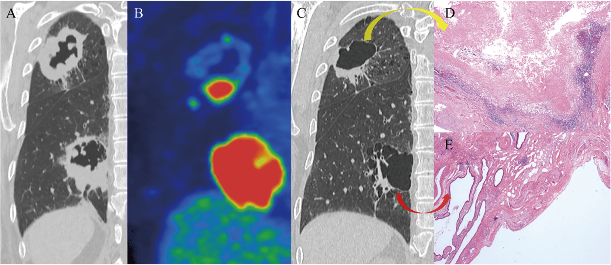
Figure 1. Simultaneous Diagnosis of Non-small Cell Lung Cancer and NTMLD. (A) Imaging detected an incidental cavitary mass in the right S2 and S6 regions. (B) Positron emission tomography/computed tomography revealed SUVmax values of 8.7 in S2 and 19 in S6. (C) Diagnosed with simultaneous cT4N0M0 stage IIIA non-small cell lung cancer (S6) and NTMLD (M. avium) (S2). After 3 months of treatment with carboplatin combined with nanoparticle albumin-bound paclitaxel, as well as rifampicin, ethambutol, clarithromycin, and amikacin, both tumors regressed. (D, E) The patient underwent right upper lobectomy and left S6 segmentectomy, achieving pathological complete remission.
Methods
Protocol and Registration
This pooled analysis was conducted and reported following the Preferred Reporting Items for Systematic Reviews and Meta-Analyses (PRISMA) guidelines.8 The study was registered with the University Hospital Medical Information Network Clinical Trials Registration (registration ID number: UMIN000056786).9
Inclusion and Exclusion Criteria
The inclusion criteria were: (1) confirmed diagnosis of lung cancer and NTMLD; (2) availability of baseline clinical characteristics (e.g., age, sex, and symptoms); (3) clinical studies, observational studies, case series, and case reports; and (4) studies in any language. The exclusion criteria were: (1) in vitro or animal experiments, reviews, meta-analyses, and duplicates; and (2) conference abstracts.
Search Strategy
We searched PubMed, CINAHL, and Igaku Chuo Zasshi for studies published between January 1, 2000, and January 31, 2025. The search terms used were: “lung cancer” and “non-tuberculosis mycobacterium” or “mycobacterium.” The quality assessment of all included studies was conducted by two reviewers (F.K. and K.Y.). Studies requiring additional evaluation were reviewed by a third reviewer (N.T.) and resolved through discussions among the three reviewers.
Definition
Lung cancer was evaluated according to the 8th edition of the TNM classification adopted by the Union for International Cancer Control.10 NTMLD was defined based on the criteria outlined in Table 3, as presented in the 2007 Official ATS/IDSA Statement.11 However, symptoms were excluded from the diagnosis due to uncertainty about which disease was responsible for the symptoms. The evaluation of cavities and lesion extent was classified based on the disease classification system of the Japan Society for Tuberculosis and Nontuberculous Mycobacteriosis.12 Furthermore, a simultaneous diagnosis was defined as cases where both conditions were confirmed within 6 months, following the definition of synchronous multiple primary malignant neoplasms.13,14
Outcomes
Given the exploratory nature of the simultaneous diagnosis of lung cancer and NTMLD, primary outcomes focused on clinical features. Statistical methods are detailed in subsequent sections.
Risk of Bias
The Newcastle-Ottawa Quality Assessment Scale (NOS) and a tool for evaluating the methodological quality of case reports and case series were used to identify the risk of bias.15 Owing to the nature of single-arm studies, case series studies (>5) received a maximum of four stars, case–control studies five stars, and cohort studies six stars. Case series (≤5) and case reports were evaluated using the methodological quality tool,16 with a maximum of five stars. Studies were included if all evaluable items in the NOS and tool received more than two stars.
Statistical Analysis
All data were analyzed using JMP Pro version 17.1.0 (SAS Institute Inc., Tokyo, Japan). Continuous variables are expressed as means and standard deviations if they follow a normal distribution; otherwise, medians and interquartile ranges (IQRs) are used. Additionally, 95% confidence intervals (CIs) were estimated. Statistical significance was set at p < 0.05. Overall survival (OS) was analyzed using the Kaplan–Meier method to estimate the median OS.
Results
Characteristics of Included Patients
A preliminary search identified 544 relevant studies from three databases (PubMed = 313, CINAHL = 25, and Igaku Chuo Zasshi = 207). After 17 duplicate records were removed, 527 studies were screened based on titles and abstracts. Among these, 485 were excluded for not meeting the inclusion criteria, primarily owing to simultaneous diagnoses of malignancies other than lung cancer (246 cases) or tuberculosis (201 cases), leaving 59 studies for eligibility assessment. Subsequently, three studies were excluded owing to insufficient information, 29 for not involving simultaneous diagnoses of lung cancer and NTMLD, and one study was withdrawn, as shown in the PRISMA flowchart (Fig. S1).
Included Studies
Finally, 26 studies17–42 involving 92 patients were analyzed. These included eight case series and 18 case reports. No phase II or III clinical trials were identified. Three studies were evaluated using the Newcastle-Ottawa Scale (NOS), receiving a four-star rating (Table S1). All analyzed case reports were judged to be included in the overall appraisal of tools for evaluating the methodological quality of case reports and case series (Table S2).
Primary Outcomes
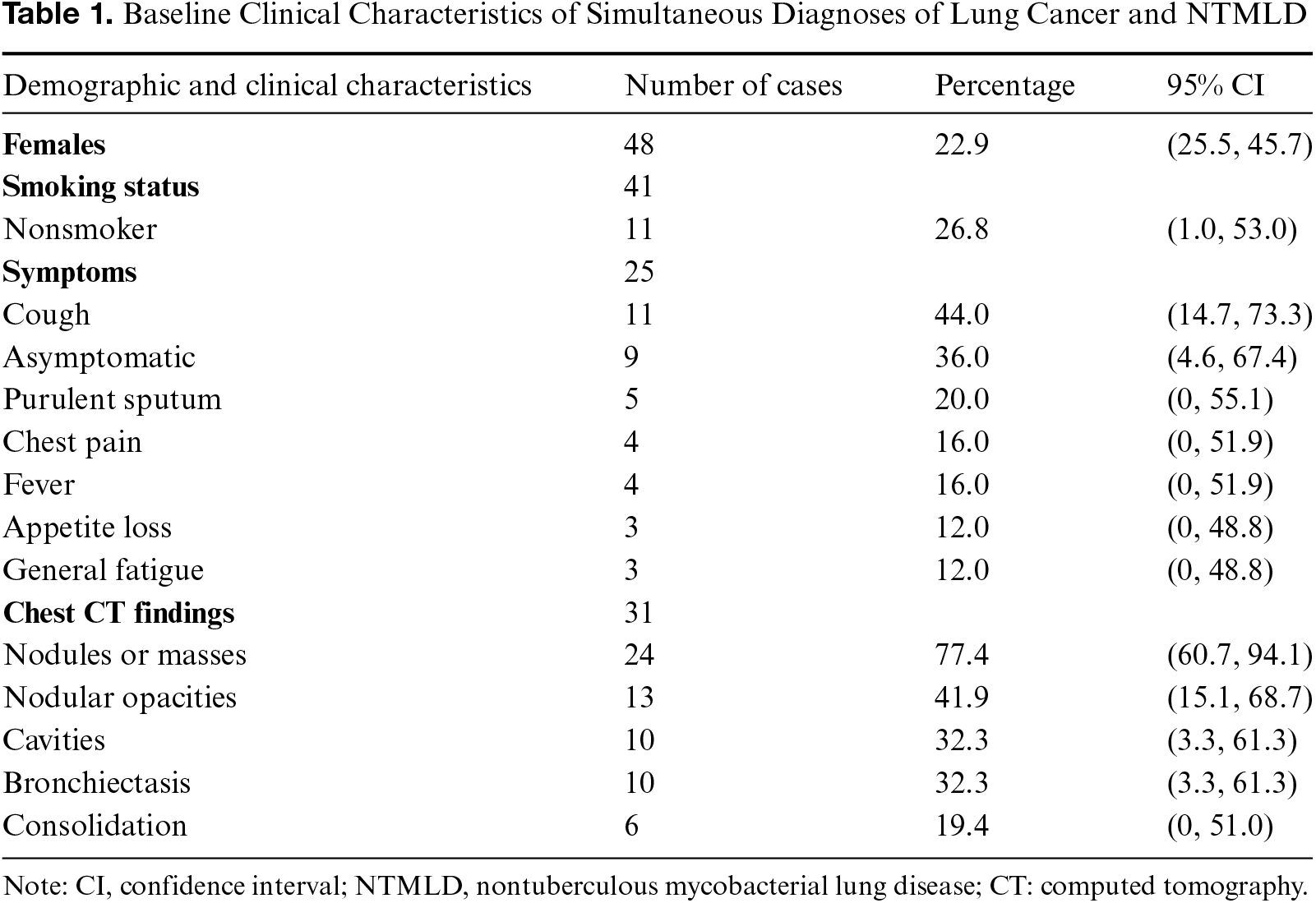
The clinical features of simultaneous diagnoses of lung cancer and NTMLD are summarized in Table 1. The mean age of patients was 69 years (IQR, 59–75). The proportion of females was significantly lower, at 22.9% (p < 0.01). Among the patients, 26.8% were nonsmokers. Common initial symptoms included cough (44.0%), purulent sputum (20.0%), and chest pain (16.0%). However, 36.0% of asymptomatic patients were found to have abnormal chest imaging findings during health checks or other medical examinations. Chest CT findings revealed nodules or masses in 77.4% of cases, followed by nodular shadows (41.9%), cavities (32.3%), bronchiectasis (32.3%), and consolidation (19.4%).
The characteristics of NTMLD and lung cancer are summarized in Table 2.
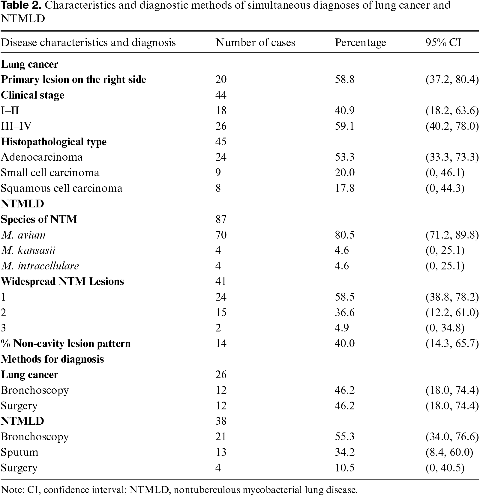
Among the cases, 58.8% of primary lung cancers were located in the right lung, 40.9% were at clinical stages I–II, and 59.1% were at stages III–IV. The histological subtypes of lung cancer were adenocarcinoma (53.3%), small cell carcinoma (20.0%), and squamous cell carcinoma (17.8%). Diagnosis of lung cancer was made by bronchoscopy (46.2%) or surgery (46.2%). No significant differences were observed in the clinical characteristics of lung cancer.
Regarding NTMLD, Mycobacterium avium was the most common pathogen (80.5%), followed by M. kansasii and M. intracellulare. Significant differences were noted between M. avium and the other species (p < 0.01). The spread of NTMLD was more commonly localized to a unilateral lung, with significantly fewer cases involving bilateral lung spread (p < 0.01). Additionally, 40.0% of NTMLD cases lacked obvious cavitary lesions. The majority of NTMLD diagnoses were made via bronchoscopy (55.3%), while 34.2% were diagnosed through sputum examination during outpatient consultations. A smaller proportion (10.5%) of cases were diagnosed through surgery. Both diseases were diagnosed almost simultaneously in 67.7% of cases, and a definitive diagnosis was made within 2 months in 87.1% of cases.
A total of 42 patients with both lung cancer and NTMLD lesions were classified into five types based on the location of the primary lesions for each disease on chest imaging (Fig. 2). Type I was defined as both lung cancer and NTMLD occurring in the same lesion. Type II was defined as both lesions located within the same lung lobe, further subclassified into Type IIa for adjacent lesions and Type IIb for nonadjacent lesions. Lesions located in different lobes of the same lung were classified as Type III, while those located in the contralateral lung were classified as Type IV. Cases with random lesion distribution or where lesion identification was unclear were categorized as Type V (unclassifiable). The distribution of cases was as follows: Type I, 16.7%; Type II, 40.5%; Type III, 21.4%; Type IV, 14.3%; and Type V, 7.1%. No significant differences in frequency were observed among the types. In histopathologic analysis, cases classified as Types I and II, and some from the random distribution pattern (Type V) showed mixed findings of lung cancer and NTMLD within the same lesion,17–19,23,24,28,30,31,36,38 whereas cases classified as Types III and IV contained only the respective lesions at the resection site.35,37
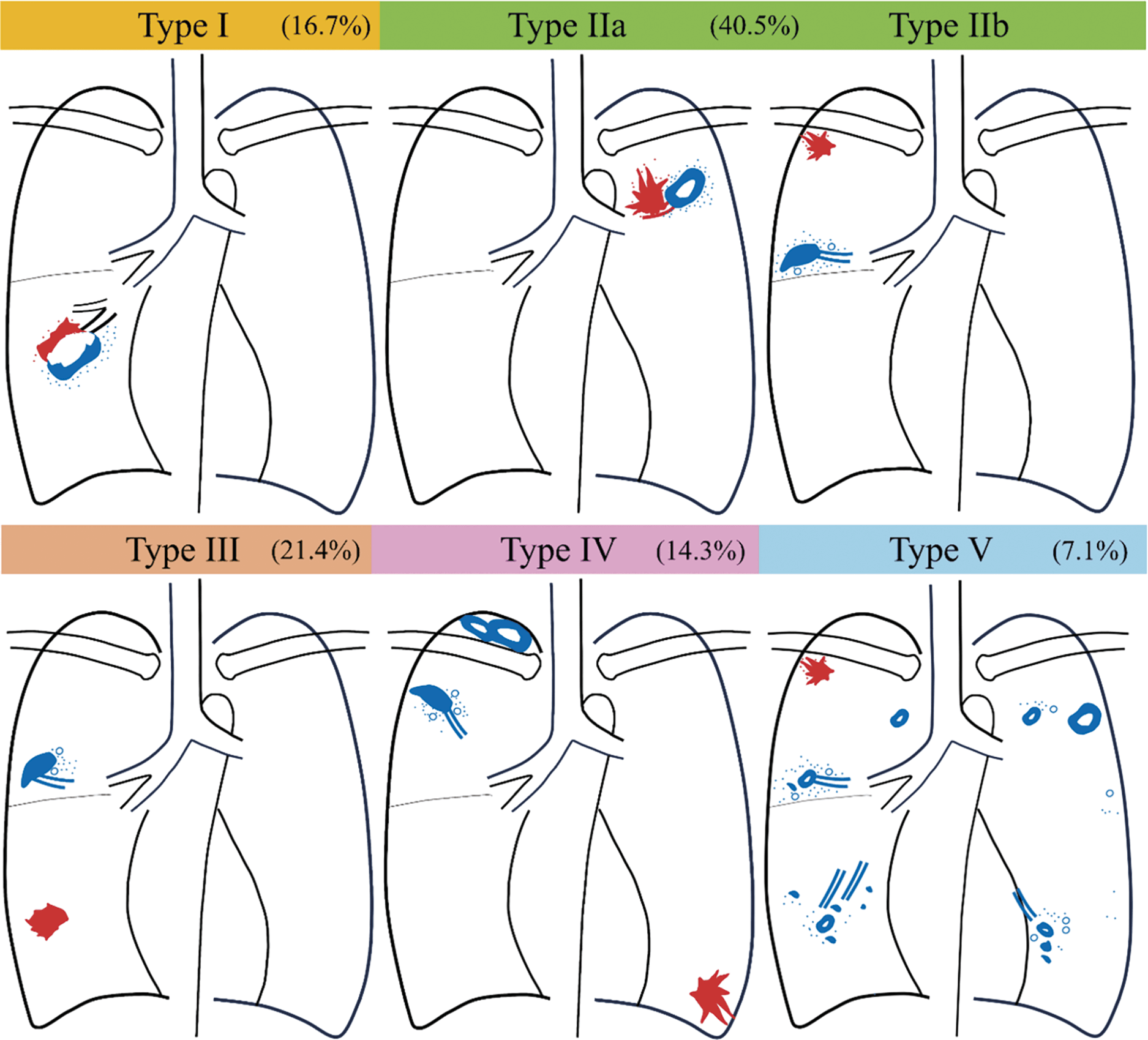
Figure 2. Classification of Lesion Distribution in Simultaneous Lung Cancer and NTMLD on Chest Imaging*. *Chest imaging, including computed tomography and plain radiography. Red lesions: Primary lung cancer; blue lesions: non-tuberculous mycobacterial lung disease; Type I: lung cancer and NTMLD present within the same lesion; Type II: both lesions located within the same lung lobe; IIa: adjacent lesions in the same lobe; IIb: nonadjacent lesions in the same lobe; Type III: lesions in different lobes of the same lung; Type IV: lesions present in the contralateral lung; Type V: unclassifiable due to random distribution or difficulty in lesion identification.
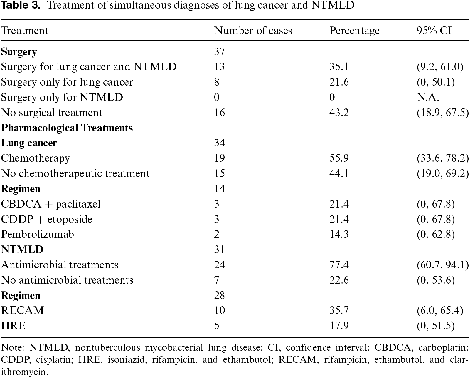
Regarding treatment, surgery was performed when complete resection was feasible based on the TNM classification of lung cancer, while no cases underwent complete resection solely for NTMLD. Chemotherapy was primarily administered in advanced-stage lung cancer cases, with some patients (7.7%) receiving programmed death-1 (PD-1) inhibitors following the approval of ICIs. Only a few studies provided details on chemotherapy regimens, but three patients receiving PD-1 inhibitors were reported in detail. Two patients had not started antimicrobial treatment before PD-1 inhibitor administration, and their NTMLD lesions (M. avium and M. fortuitum) worsened afterward. They then initiated antimicrobial treatment, continued the PD-1 inhibitor regimen, and showed improvement in NTMLD lesions.39,40 Antimicrobial treatment for NTMLD was significantly more common (p < 0.01), with RECAM being the most frequently used regimen. Among the 22 patients with available OS data, the median OS was 23 months.
Discussion
This study describes the clinical characteristics of cases with simultaneous diagnoses of lung cancer and NTMLD since 2000, a period marked by the widespread adoption of chest CT scans. The majority of cases were male smokers, typically identified on chest imaging when patients presented with symptoms such as cough, sputum, or chest pain, or during health checks and other medical examinations. M. avium was the most frequently diagnosed NTMLD species, often localized to one lung. Adenocarcinoma was the most common histologic type of lung cancer, with no significant differences observed. Furthermore, we categorized patients with simultaneous diagnoses of lung cancer and NTMLD into five major types based on the locations of their primary lesions.
Several reports have shown that the rate of lung cancer complications in patients with nontuberculous mycobacterial disease is higher than in healthy individuals, but the findings remain controversial.43 These studies have not examined the relationship between the locations of the lesions for these two diseases. Calculations based on Tamura et al.’s report indicate that the rate of simultaneous diagnosis was 1.8% for NTMLD patients with lung cancer and 1.0% for lung cancer patients with NTMLD as a complication.20 They reported that half of these patients had the primary lesions of both diseases within the same lung lobe.20
The lung parenchyma in patients with lung cancer is often compromised by a combination of cancer cell metastasis and damage caused by surgery, radiation, and antineoplastic drugs, creating a predisposition to infection.44 Additionally, the use of cytotoxic antineoplastic drugs and cancer-induced cachexia contributes to decreased body mass index and the development of sarcopenia, leading to reduced interleukin-15 production—essential for T cell proliferation—which in turn results in increased PD-1 expression and diminished T cell function, and further progression of infections.2 Infections are more likely to develop in the respiratory system when physiological, immunological, or morphological abnormalities are present, along with impaired drainage.20,23 These factors may contribute to the occurrence of NTMLD near lung cancer.
In the present study, Type I cases were defined as those with NTM lesions coexisting within the cavities formed by lung cancer. In Type IIa cases, where the lesions were adjacent to each other, peripheral drainage function was impaired by central lung cancer lesions, leading to frequent NTMLD complications at the peripheral sites of lung cancer or adjacent lung cancer lesions owing to chronic NTMLD. These types constituted nearly 60% of the cases, suggesting an interplay between immunity and inflammation in the coexistence of both diseases. However, in cases where the lesions of both diseases were distant from each other, such as in Type III and Type IV, the pathological findings of each disease were similar to those observed in noncomplicated cases, especially in cases like Type IV, which accounted for 14.3% of the total. The association between lesion locations in both diseases by type may be linked to disease progression and occasional complications.
There are several limitations to this study. First, all the enrolled studies were retrospective analyses. Second, the data were primarily derived from case reports, which are susceptible to publication bias and do not permit sensitivity analysis. Therefore, statistical significance was interpreted as a reference value. Third, the analysis of symptoms in NTMLD diagnosis was waived owing to the overlap with lung cancer. Fourth, NTMLD is caused by M. avium, the predominant species, which may explain the higher number of studies from Japan. However, this study enabled us to classify lung cancer and NTMLD into five categories based on the relationship between primary lesion locations. In cases where lung cancer and NTMLD coexist, further insights may be gained by examining the spatial relationship between their primary lesions.
Conclusions
Simultaneous diagnoses of lung cancer and NTMLD were often made incidentally. In approximately half of the cases, both lesions were located in the same lung lobe, while in some instances, they were found in the contralateral lung according to our classification. However, the small sample size and the retrospective nature of the enrolled studies introduced significant bias, emphasizing the need for further research to validate this classification.
Acknowledgments
None.
Funding Source
None.
Author Contributions
F.K. and K.Y. interpreted the data and drafted the original and revised manuscript. These two authors contributed equally to this work. N.T. and Y.K. contributed to the analysis and data curation. S.S., R. Orii, R. Osada, M.K., A.K., and H.O. were responsible for data curation. H.K. and K.T. made substantial contributions to revising the manuscript drafts. All authors have reviewed and approved the final version of the manuscript and agree to be accountable for their respective contributions to the work.
Data Availability
The datasets used in the current study are available from the corresponding author upon reasonable request.
Ethical Statement
The article does not involve the participation of any animals. The patient provided written informed consent for the publication of this report and accompanying images.
Conflict of Interest
F.K. serves as a member of the editorial board for the Journal of Clinical Question.
Supplemental Information
Supplemental information for this article can be found online at https://sup.jclinque.com/api/articles/59/download-suppl.
References
[1] World Health Organization (Lung Cancer). https://www.who.int/news-room/fact-sheets/detail/lung-cancer. Published 2024, Accessed on January 30, 2025.
[2] Vaddi A, Hulsebus HJ, O'Neill EL, et al. A narrative review of the controversy on the risk of mycobacterial infections with immune checkpoint inhibitor use: does Goldilocks have the answer? J Thorac Dis. February 2024;29(2):1601–1624. doi:10.21037/jtd-23-1395.
[3] Georgakopoulou VE, Garmpis N, Mermigkis D, et al. Pulmonary adverse events due to immune checkpoint inhibitors: a literature review. Monaldi Arch Chest Dis. October 11, 2021;92(2). doi:10.4081/monaldi.2021.2008.
[4] Forder A, Zhuang R, Souza VGP, et al. Mechanisms contributing to the comorbidity of COPD and lung cancer. Int J Mol Sci. February 2, 2023;24(3). doi:10.3390/ijms24032859.
[5] Tzouvelekis A, Gomatou G, Bouros E, Trigidou R, Tzilas V, Bouros D. Common pathogenic mechanisms between idiopathic pulmonary fibrosis and lung cancer. Chest. August 2019;156(2):383–391. doi:10.1016/j.chest.2019.04.114.
[6] Yu YH, Liao CC, Hsu WH, et al. Increased lung cancer risk among patients with pulmonary tuberculosis: a population cohort study. J Thorac Oncol. January 2011;6(1):32–37. doi:10.1097/JTO.0b013e3181fb4fcc.
[7] Morimoto K, Hamashima R, Yamada T, et al. Clinical significance of chronic pulmonary aspergillosis in lung cancer patients undergoing anticancer drug therapy. Thorac Cancer. September 2024;15(26):1882–1888. doi:10.1111/1759-7714.15416.
[8] Page MJ, McKenzie JE, Bossuyt PM, et al. Mapping of reporting guidance for systematic reviews and meta-analyses generated a comprehensive item bank for future reporting guidelines. J Clin Epidemiol. February 2020;118(7):60–68. doi:10.1016/j.jclinepi.2019.11.010.
[9] University Hospital Medical Information Network. https://center6.umin.ac.jp/cgi-open-bin/ctr/ctr_view.cgi?recptno=R000064907. Published 2024, Accessed on January 30, 2025.
[10] Brierly JDGM, Wittekind C, eds. UICC TNM Classification of Malignant Tumours. 8th ed. Wiley Blackwell; 2017.
[11] Griffith DE, Aksamit T, Brown-Elliott BA, et al. An official ATS/IDSA statement: diagnosis, treatment, and prevention of nontuberculous mycobacterial diseases. Am J Respir Crit Care Med. February 2007;15(4):367–416. doi:10.1164/rccm.200604-571ST.
[12] Takase A. X-ray classification of pathological types of tuberculosis. Kekkaku. June 2011;86(6):607–617.
[13] Warren S, Gates O. Multiple primary malignant tumors: a survey of the literature and statistical study. Am J Cancer. 1932;16:1358–1414.
[14] Coyte A, Morrison DS, McLoone P. Second primary cancer risk—the impact of applying different definitions of multiple primaries: results from a retrospective population-based cancer registry study. BMC Cancer. April 18, 2014;14(1):272. doi:10.1186/1471-2407-14-272.
[15] Stang A. Critical evaluation of the Newcastle-Ottawa scale for the assessment of the quality of nonrandomized studies in meta-analyses. Eur J Epidemiol. September 2010;25(9):603–605. doi:10.1007/s10654-010-9491-z.
[16] Murad MH, Sultan S, Haffar S, Bazerbachi F. Methodological quality and synthesis of case series and case reports. BMJ Evid Based Med. April 2018;23(2):60–63. doi:10.1136/bmjebm-2017-110853.
[17] Souilamas R, Danel C, Chauffour X, Riquet M. Lung cancer occurring with Mycobacterium xenopi and Aspergillus. Eur J Cardiothorac Surg. Jul 2001;20(1):211–213. doi:10.1016/S1010-7940(01)00720-5.
[18] Fujiwara K, Yasumitsu T, Nakagawa K, et al. A case of nontuberculous mycobacteriosis and an adenocarcinoma in a single lesion in the lung. Jpn J Chest Dis. May 2002;61(5):470–473.
[19] Kobayashi K, Yano S, Kato K, et al. A case of M. fortuitum lung disease with small-cell lung cancer. J Jpn Respir Soc. May 2004;42(5):424–428.
[20] Tamura A, Hebisawa A, Sagara Y, et al. Pulmonary nontuberculous mycobacteriosis in patients with lung cancer. Kekkaku. June 2004;79(6):367–373.
[21] Tsunezuka Y, Ishikawa N, Hiranuma C, Sato H, Oda M, Watanabe G. Mycobacteria other than tuberculosis combined primary lung cancer. Kyobu Geka. February 2004;57(2):119–122.
[22] Yonemori K, Takeda Y, Toyota E, Kobayashi N, Kudo K. Potential interactions between irinotecan and rifampin in a patient with small-cell lung cancer. Int J Clin Oncol. June 2004;9(3):206–209. doi:10.1007/s10147-004-0394-4.
[23] Tamura A, Hebisawa A, Sagara Y, et al. Coexistence of lung cancer and active pulmonary mycobacteriosis. Kekkaku. May 2005;80(5):413–419.
[24] Tokushima T, Araki K, Metsugi H, et al. Three cases of lung cancer inpatients with pulmonary nontuberculous mycobacteriosis. J Jpn Assoc Chest Surg. January 2006;20(1):33–39. doi:10.2995/jacsurg.20.33.
[25] Abe K, Yamazato M, Ohtani S, et al. Peculiar “chestnuts in burrs” formation in MGIT cultures of pulmonary Mycobacterium xenopi cases. Rinsho Byori. December 2007;55(12):1080–1083.
[26] Mori M, Fushitani K, Nakazawa Y, et al. Two cases of non-small cell lung cancer complicated with non-tuberculous mycobacterium (NTM) infection successfully treated with anti-NTM agents and anti-cancer agents. Jpn J Chest Dis. July 2008;67(7):613–621.
[27] Matsumoto A, Enomoto T, Muroya Y, et al. Pulmonary non-tuberculous mycobacteriosis (Mycobacterium intracellulare) with cavities developing in a non-small cell lung cancer patient during chemotherapy. J Jpn Respir Soc. August 2010;48(8):609–613.
[28] Koga T, Sato R, Kamimura T, Nishimura M, Kage M, Matsuo K. Concurrent lung cancer in non-tuberculous mycobacteriosis: case report. Kurume Med J. November 2011;58(3):87–90. doi:10.2739/kurumemedj.58.87.
[29] Lande L, Peterson DD, Gogoi R, et al. Association between pulmonary mycobacterium avium complex infection and lung cancer. J Thorac Oncol. 2012;7(9):1345–1351. doi:10.1097/JTO.0b013e31825abd49.
[30] Ota H, Kawai H. Coexisting pulmonary mycobacterium avium intracellulare complex (MAC) disease and lung adenocarcinoma in a solitary pulmonary nodule. J Jpn Assoc Chest Surg. May 2012;26(4):481–485. doi:10.2995/jacsurg.26.481.
[31] Ozeki N, Fukui T, Niwa Y, Yoshida K, Yatabe Y, Mitsudomi T. Lung adenocarcinoma coexistent with pulmonary non-tuberculous mycobacteriosis. Jpn J Lung Cancer. April 2012;52(2):238–241.
[32] Hosoda C, Hagiwara E, Shinohara T, et al. Clinical characteristics of pulmonary Mycobacterium avium complex infection complicated with lung cancer. Kekkaku. August 2014;89(8):691–695.
[33] Tashiro H, Takahashi K, Kato G, Komiya K, Aragane-Sueoka N, Hayashi S. A case of pulmonary infection with Mycobacterium fortuitum during chemotherapy for lung cancer. J Jpn Respir Soc. May 2014;3(3):438–441.
[34] Tsuji T, Tsuyuguchi K, Tachibana K, et al. Analysis of the impact of lung cancer treatment on nontuberculous mycobacterial lung diseases. Respir Investig. January 2017;55(1):45–50.
[35] Tsushima Y, Kawakami H, Ishimori S, Anakura Y. A case of primary lung cancer concomitant with pulmonary non-tuberculous mycobacteriosis in the ipsilateral lobe. J Jpn Assoc Chest Surg. March 2017;31(2):227–232.
[36] Taira N, Kawasaki H, Takahara S, Chibana K, Atsumi E, Kawabata T. The presence of coexisting lung cancer and non-tuberculous mycobacterium in a solitary mass. Am J Case Rep. June 26, 2018;19:748–751. doi:10.12659/ajcr.908090.
[37] Komiya K, Kuwahara M, Yamamoto S, et al. A case of right upper lobe lung adenocarcinoma diagnosed concurrently with non-tuberculous mycobacterial (NTM) infection. Jpn J Clin Exp Med. July 2019;96(7):839–842.
[38] Walker CM, Zhou CY, Pathak V. Adenocarcinoma of the lung with concurrent Mycobacterium avium complex infection. Lung India. July–August 2021;38(4):365–367. doi:10.4103/lungindia.lungindia_470_19.
[39] Hirabayashi T, Sonehara K, Nozawa S, Ikuyama Y, Komatsu M, Hanaoka M. A case of lung cancer with exacerbated Mycobacterium avium lung disease under pembrolizumab treatment. J Jpn Respir Soc. September 2022;11(5):301–304.
[40] Zhang CC, Chen P. Anti-programmed death 1 antibody in the treatment of coexistent Mycobacterium fortuitum and lung cancer: a case report. World J Clin Cases. April 2022;26(12):3801–3807. doi:10.12998/wjcc.v10.i12.3801.
[41] Agarwal S, Nair G, Patel SM, Deshpande P, Sarangdhar N. A rare case of co-existing non-small cell lung carcinoma and non-tuberculous mycobacteria. Cureus. December 2023;15(12):e50456. doi:10.7759/cureus.50456.
[42] Mishra P, Kondisetti M, Patil A, Sarangdhar N. Pulmonary mycobacterium avium complex with adenocarcinoma of the lung: a case report. Cureus. August 2024;16(8):e66589. doi:10.7759/cureus.66589.
[43] Kusumoto T, Asakura T, Suzuki S, et al. Development of lung cancer in patients with nontuberculous mycobacterial lung disease. Respir Investig. March 2019;57(2):157–164. doi:10.1016/j.resinv.2018.11.004.
[44] Daley CL, Iseman M. Mycobacterium avium complex and lung cancer: chicken or egg? Both? J Thorac Oncol. September 2012;7(9):1329–1330. doi:10.1097/JTO.0b013e318265a7ef.
 |
Copyright: © 2025 Kashizaki et al. This work is licensed under a Creative Commons Attribution 4.0 International License, which permits unrestricted use, distribution, and reproduction in any medium, provided the original work is properly cited. |