| Journal of Clinical Question, 2024, Vol. 1, No. 2, 12–23 https://doi.org/10.69854/jcq.2024.0003 Advance access publication date 03 September 2024 |
 |
Meta-Analysis
Clinical Features of Pneumocystis Pneumonia in Non-Human Immunodeficiency Virus-Infected Patients: A Systemic Review and Meta-Analysis
Division of Oncology, Department of Internal Medicine, Teikyo University School of Medicine, Tokyo, Japan.
*Corresponding Author: e-mail: nseki@med.teikyo-u.ac.jp
Submitted: August 06, 2024 Accepted: September 02, 2024
Clinical Question Box
What are the clinical features of pneumocystis pneumonia in non-human immunodeficiency virus-infected patients?
Pneumocystis pneumonia was more likely to occur in men than in women at the age of 60, with a sex ratio of about 3 to 2 (males to females). The main symptoms were dyspnea and fever, which were observed in >70% of patients, accompanied by cough in >50% of patients. Ground glass opacity might offer a clue to diagnosing pneumocystis pneumonia in high-risk patients.
Abstract
Objectives: Pneumocystis pneumonia (PCP) is an opportunistic disease that causes potentially fatal pneumonia in immunocompromised individuals. The clinical features of PCP without HIV remain incompletely understood. Methods: This study aimed to identify the clinical features of PCP without HIV in a systematic review following a meta-analysis. Results: 65 articles that included 10,133 PCP patients without HIV infection were enrolled. PCP occurred most commonly at age 59.2 years (95% CI: 57.7, 60.7) in a gender ratio of approximately 3 to 2 (males to females). Dyspnea, fever, cough, and sputum were nonspecific clinical findings in 73% (95% CI: 69, 79), 73% (95% CI: 65, 81), 56% (95% CI: 48%, 64%), and 32% (96%CI: 16, 48) of patients, respectively. Viral, bacterial, and fungal co-infection were observed in 28% (95% CI: 13, 44), 19% (95% CI: 13, 25), and 11% (95% CI: 6, 16) of patients, respectively. Laboratory data showed a trend of elevated WBC, LDH, CRP, β-D glucan, and KL-6. Ground glass opacity (GGO) was found in 87% (95% CI: 83, 91) of patients. In-hospital mortality was 41% (95% CI: 35, 46). Conclusions: PCP is a life-threatening disease in immune-compromised patients. Despite being a nonspecific clinical finding, GGO might offer a clue to diagnosing PCP in high-risk patients.
Keywords: Pneumocystis pneumonia, clinical features, non-HIV, ground glass opacity.
Introduction
In individuals who are immunocompromised, the opportunistic fungal pathogen Pneumocystis jirovecii is capable of causing life-threatening pneumonia as the causative agent of pneumocystis pneumonia (PCP).1 Human immunodeficiency virus (HIV)-infected patients are at the highest risk of PCP; however, non-HIV-infected immunocompromised patients, including those with chronic corticosteroid use, hematological or solid malignancies, organ transplantation, and immunomodulatory or biological therapy have a substantial risk of developing PCP.2,3
HIV-infected patients usually develop a subacute course of the disease, whereas respiratory insufficiency is usually more severe in non-HIV patients than in the HIV-infected population. Pneumocystis is more challenging to detect in non-HIV patients because of the smaller numbers of organisms in the lungs.4 In addition, the mortality rate is 30%–50% in patients without HIV and 10%–12% in patients with HIV.5 The diagnosis of PCP relies on a critical evaluation of clinical symptoms, risk factors, radiologic features, and microbiological tests. Computed tomography (CT) examination is helpful if there is clinical suspicion of PCP.
A clear overview of PCP in non-HIV patients is essential for diagnostic progress and treatment in clinical practice; however, this is currently lacking, and no meta-analysis has appraised the clinical features in this group. Therefore, we conducted a systematic review and meta-analysis to explore the general characteristics, comorbidity, co-infection, and laboratory and radiographic findings of PCP obtained in observational studies of non-HIV patients.
Methods
Study Overview
The protocol of this systematic review and meta-analysis followed the guidelines of the Preferred Reporting Items for Systematic Reviews and Meta-Analyses (PRISMA) statement. It was registered on the website of the University Hospital Medical Information Network Clinical Trials Registration (UMIN 000046230).6,7 Because of the nature of the study, Institutional Review Board approval was not required.
Study Search
Four major online databases (PubMed, Web of Science, Cochrane, and Embase) were searched from January 1, 2000 to October 30, 2022. The following search strategy was used for PubMed: (((((pneumocystis pneumonia) OR (pneumocystis jirovecii)) OR (pneumocystis infections)) OR (pneumocystis carinii)) AND (((((non-HIV) OR (non-AIDS)) OR (non-HIV-infected)) OR (HIV-uninfected)) OR (HIV-negative))) AND (((symptom) OR (clinical feature)) OR (diagnosis)). Two authors (H.C. and M.I.) independently screened the titles and abstracts and carefully evaluated the full text to select eligible articles; in cases of discrepancy, a consensus was reached through discussion. Review articles and the included original articles were hand-searched (H.C. and M.I.) for additional research papers that met the inclusion criteria.
Inclusion and Exclusion Criteria
Full-length articles published in any language that provided data regarding the clinical features of PCP were included. The inclusion criteria were: (1) patients with PCP, (2) patients without HIV, and (3) age ≥16 years. The exclusion criteria were: (1) detailed data not available, (2) fewer than 10 cases, and (3) limited information regarding clinical features.
Risk of Bias
Two reviewers independently assessed the methodological quality of the selected studies using the Newcastle–Ottawa quality assessment to evaluate the quality of observational studies.8 Due to the nature of single-arm studies, the “selection of the non-exposed cohort” and “comparability” domains were not applicable in this study. The modified Newcastle–Ottawa quality assessment scored six stars. Any disagreement between the reviewers was discussed, and agreement was reached by consensus.9
Outcomes
The clinical features of PCP were appraised in terms of the following five aspects: general characteristics, laboratory findings, comorbidity, existence of other microorganisms, and radiographic findings. General characteristics included age, sex, body mass index (BMI), prophylaxis, time from symptom onset to treatment, and hospital mortality. The following laboratory findings were collected: white blood cells (WBC), lactase dehydrogenase (LDH), albumin, c-reactive protein (CRP), β-D glucan, and sialylated carbohydrate antigen 6 (KL-6). Comorbidities included hematologic malignancies, solid tumors, immunosuppression (immunosuppressive drugs for nonmalignant diseases), and any transplant. Symptoms included dyspnea, fever, cough, and sputum. Although it is difficult to distinguish between co-infection and detection of the existence of other microorganisms, most previous studies have reported co-infection. Co-infection was classified as bacterial, viral, cytomegalovirus (CMV), and fungal. The radiographic findings were classified as ground glass opacity (GGO), consolidation, lung nodules, septal thickening, pleural effusion, and pneumothorax.
Data Extraction and Statistics
Two authors (M.I. and S.T.) independently extracted the following data from the studies: first author, publication year, country of publication, number of patients with PCP, clinical features, and Newcastle–Ottawa quality assessment-related information. All analyses were performed using Review Manager software version 5.3 (Cochrane Collaboration, Oxford, UK). Figures illustrated using Review Manager were adjusted as necessary. Heterogeneity was evaluated with the I2 statistic and interpreted as follows: I2 = 0%, no heterogeneity; 0% < I2 < 25%, the least heterogeneity; 25% ≤ I2 < 50%, mild heterogeneity; 50% ≤ I2 < 75%, moderate heterogeneity; and 75% ≤ I2, strong heterogeneity.10 A p-value of <0.05 was considered significant.
Results
Study Search and Study Characteristics
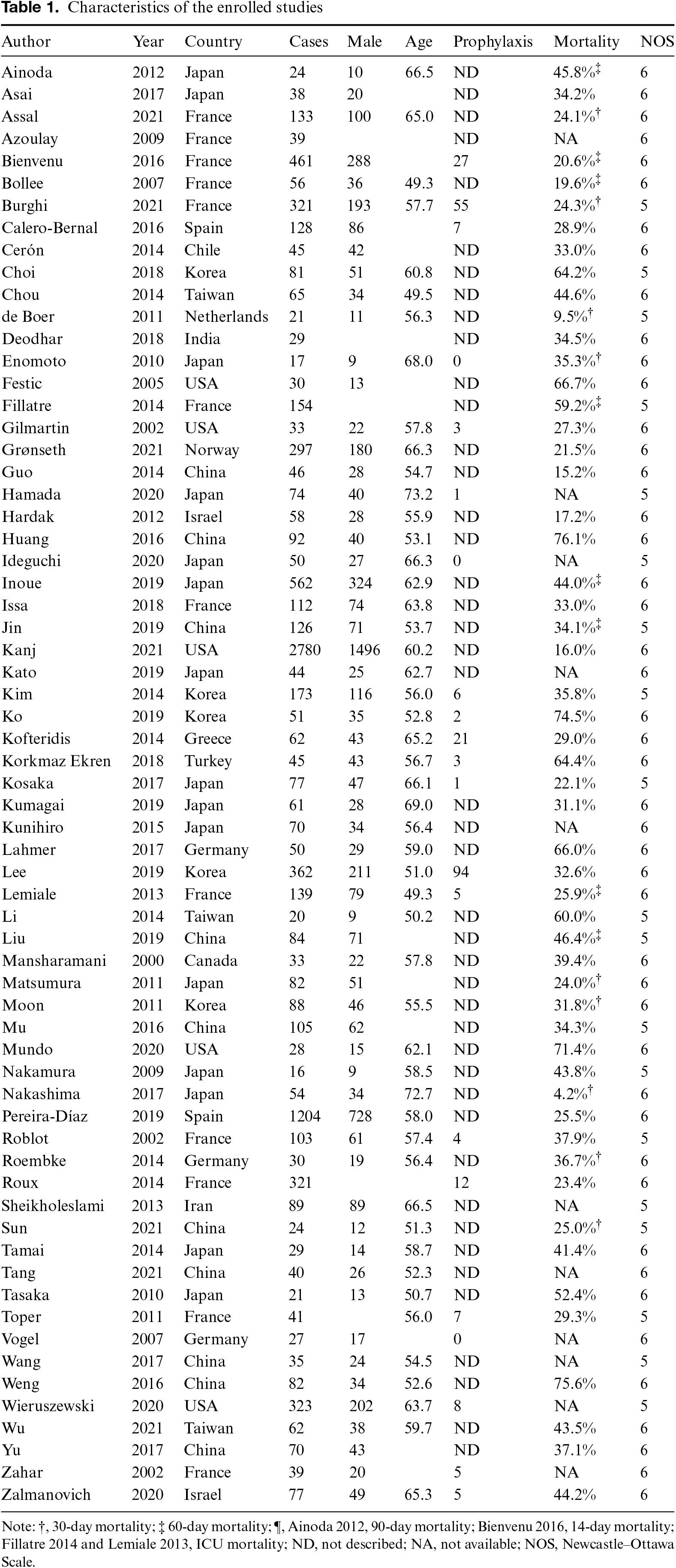
A total of 581 articles were identified, including 578 articles by database search and three articles by hand search. Of the 581 articles, 416, 179, and 65 remained after removing duplication, screening for eligibility, and full-length article reading, respectively (Fig. S1). The analysis included 65 retrospective studies (Table 1).11–75 All but two were written in English.19,32 Of the 65 reports, 15 were from Japan, 13 were from China, 12 were from France, 5 were from Korea and the USA, 3 were from Germany, and 2 were from Israel and Spain. These studies included a total of 10,133 PCP patients without HIV infection. Among the studies, the modified Newcastle–Ottawa scale ranged from 5 to 6 stars. The method for diagnosis of PCP showed some variation among studies (Table S1). Polymerase chain reaction (PCR) was conducted in 43 studies. Clinical and/or radiological findings were also included as diagnostic criteria for PCP in 39 studies. Elevation of β-D-glucan was considered necessary in 6 studies, and no diagnostic criteria were provided in 5 studies.
General Characteristics and Laboratory Findings
Table 2 lists the general characteristics of patients with PCP without HIV. In an analysis of 50 studies, the mean age was 59.2 years (95% confidence interval (CI): 57.7, 60.7; I2 = 95%; Fig. S2). Males comprised 60% of patients (95% CI: 58, 63; I2 = 85%; Fig. S3). Mean BMI was 22.1 kg/m2 (95% CI: 20.0, 24.1; I2 = 0%; Fig. S4), and 8% of patients underwent prophylaxis for PCP (95% CI: 5, 11; I2 = 92%; Fig. S5). Trimethoprim/sulfamethoxazole was used as prophylaxis in 11 studies. However, detailed information should have been provided. The other eight studies (Table S2) did not state the length of prophylaxis. The mean time from symptom onset to treatment was 8.9 days (95% CI: 6.8, 10.9; I2 = 94%; Fig. S6), and mean in-hospital mortality due to PCP was 41% (95% CI: 35, 46; I2 = 95%; Fig. S7) in 34 studies.
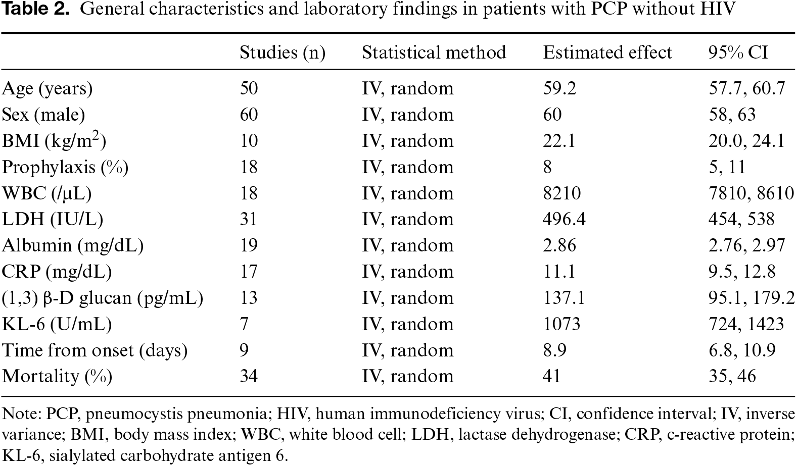
The laboratory findings revealed elevated WBC, LDH, CRP, β-D glucan, and KL-6 and a decreased albumin value in the overall patients. The average values were as follows: WBC, 8,210/μL (95% CI: 7,810, 8,610; I2 = 52%; Fig. S8); LDH, 496 IU/L (95% CI: 454, 538; I2 = 94%; Fig. S9); albumin, 2.86 mg/dL (95% CI: 2.76, 2.97; I2 = 91%; Fig. S10); CRP, 11.1 mg/dL (95% CI: 9.5, 12.8; I2 = 89%; Fig. S11); β-D glucan, 137.1 pg/dL (95% CI: 95.1, 179.2; I2 = 100%; Fig. S12); and KL-6, 1073 (95% CI: 724, 1423; I2 = 91%; Fig. S13).
Comorbidity
The frequencies of comorbidities in patients with PCP without HIV infection are listed in Table S1. The most frequent comorbidities were hematologic malignancies and use of immunosuppressive drugs for nonmalignant diseases, followed by transplant and solid tumor, as follows: 32% (95% CI: 27, 36; I2 = 97%; Fig. S14); 32% (95% CI: 27, 36; I2 = 96%; Fig. S15), 19% (95% CI: 16, 23; I2 = 95%; Fig. S16), and 16% (95% CI: 14, 18; I2 = 81%; Fig. S17).
Symptoms
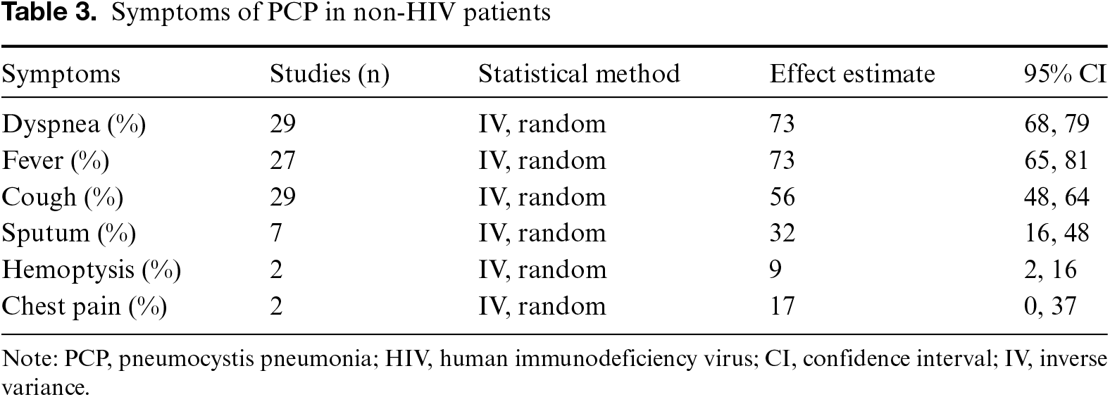
Table 3 lists the symptoms reported in at least two studies. The most frequently reported symptoms were dyspnea and fever, followed by cough, sputum, hemoptysis and chest pain, as follows: 73% (95% CI: 68, 79; I2 = 94%; Fig. S18), 73% (95% CI: 65, 81; I2 = 97%; Fig. S19), 56% (95% CI: 48, 64; I2 = 96%; Fig. S20), 32% (95% CI: 16, 48; I2 = 95%; Fig. S21), 9% (95% CI: 2, 16; I2 = 49%; Fig. S22), and 17% (95% CI: 0, 37; I2 = 85%; Fig. S23), respectively. The meta-analysis did not include articles that reported limited fatigue and body-weight loss data.
Co-infection
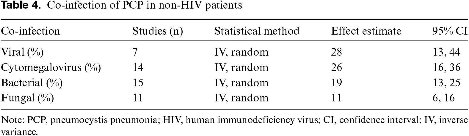
Table 4 lists data regarding the detected microorganisms. In patients with PCP without HIV infection, viral microorganisms were detected most frequently, followed by bacterial and fungal, as follows: 28% (95% CI: 13, 44; I2 = 97%; Fig. S24), 19% (95% CI: 13, 25; I2 = 89%; Fig. S25), and 11% (95% CI: 13, 44; I2 = 97%; Fig. S26), respectively. Cytomegalovirus (CMV) was observed in 26% (95% CI: 16, 36; I2 = 97%; Fig. S27) of PCP patients. The methods used to identify CMV, viruses, and bacteria are shown in Tables S3, S4, and S5, respectively. A total of 14 studies reported a positive result for CMV, but only eight described the identification method. Among these, CMV antigenemia assay and real-time PCR were used in three studies each. Positive results were found for CMV in 25.5% and 47.3% of antigenemia assay and real-time PCR, respectively. The co-infection of viruses and bacteria was reported in 7 and 15 studies, respectively. Microbiological confirmation was used as the detection method in most of these studies.
Radiographic Findings
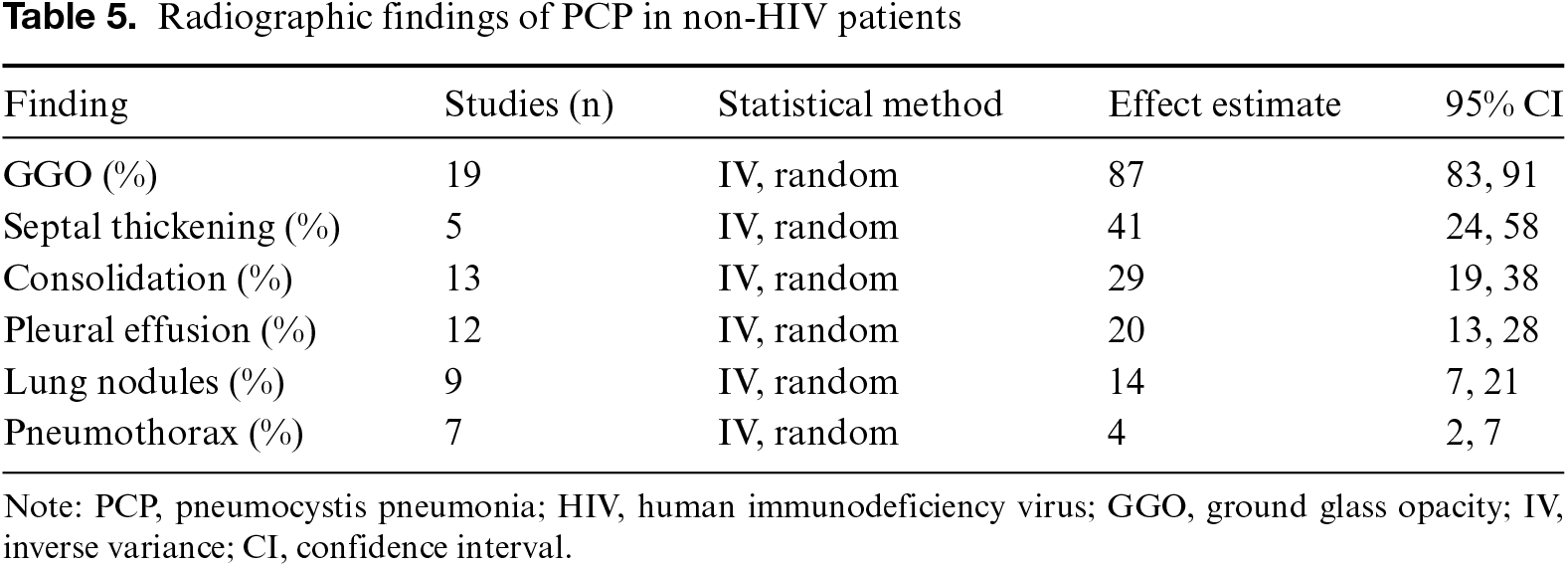
Table 5 summarizes the radiographic findings. GGO was most the frequent radiographic finding, followed by septal thickening, consolidation, pleural effusion, lung nodules, and pneumothorax, as follows: 87% (95% CI: 83, 91; I2 = 95%; Fig. S28), 41% (95% CI: 24, 58; I2 = 91%; Fig. S29), 29% (95% CI: 19, 38; I2 = 94%; Fig. S30), 20% (95% CI: 13, 28; I2 = 90%; Fig. S31), 14% (95% CI: 7, 21; I2 = 91%; Fig. S32), and 4% (95% CI: 2, 7; I2 = 63%; Fig. S33), respectively.
Discussion
The general features of PCP, including clinical presentation and laboratory and radiographic findings, were identified after analysis of 10,133 cases of PCP in patients without HIV. PCP was more likely to occur in men than in women at the age of 60 years, with a sex ratio of about 3 to 2 (males to females). The main symptoms were dyspnea and fever, which were observed in >70% of patients, accompanied by cough in >50% of patients. Most studies did not specify the type of cough; however, cough was accompanied by sputum in >30% of patients. The co-infection was frequently observed, and CMV was detected in >25% of patients. The laboratory findings revealed decreased albumin but no apparent weight loss in PCP patients. Elevated WBC, LDH, CRP, β-D glucan, and KL-6 were also observed in these patients. More than 60% of patients had a comorbidity of either hematologic malignancy or immunosuppressive drug use for nonmalignant disease. Eight percent of PCP patients underwent prophylaxis for PCP, and in-hospital mortality was high (~41%), which is in agreement with the results of previous studies.3,34,76
PCP is of increasing importance in non-HIV immunocompromised patients who present with severe respiratory distress and low fungal loads. β-D glucan level was found to be very useful in the diagnosis of PCP, and seven studies included β-D glucan positivity as a condition for the diagnosis of PCP. In contrast, three studies have reported negative β-D glucan in non-HIV patients, ranging from 12% to 27.2%.38,52,56 These reports were all from Japan, which suggests possible racial differences or differences in laboratory examination methods between countries. The high rate of CMV positivity was surprising. CMV generally produces an asymptomatic or minimally symptomatic acute illness in immunocompetent patients. Detecting viral DNA/RNA does not necessarily mean clinically relevant disease but may also indicate viral ‘dead’ material.
Most importantly, CMV reactivation can be found in sick airways, and low levels of CMV can be found in peripheral blood. However, the enrolled articles provided limited information that would enable differences to be identified between genetic information and CMV infection. The exact proportion of patients with the existence of CMV and how CMV infection affects the clinical progress of PCP in non-HIV patients remains unclear.
GGO was observed in almost 90% of PCP patients. Given the nonspecific nature of the clinical findings, this radiology finding appears to be an essential clue for confirming a diagnosis of PCP, even in asymptomatic high-risk patients. Numerous studies reported less common radiographic findings such as consolidation, pleural effusion, and lung nodules. Combining co-infection and PCP might be necessary to interpret the various image findings. The diagnostic accuracy of radiology in PCP remains unclear, and further studies are required.
PCP is challenging to diagnose. Asymptomatic lung colonization can occur in people with normal immune systems who may unknowingly become reservoirs (asymptomatic carriers) for the spread of pneumocystis to immunocompromised individuals.77 A few articles have discussed the relevance of sputum colonization vs. bronchoalveolar lavage sampling and the sensitivities between PCR and microscopy staining.
There were several limitations in this study. First, high heterogeneity was observed in almost all of the analyses performed due to the observational nature of the studies and their differing purposes. Although random effects were considered in data analysis, selection bias was possible. Second, the detection of pathogens is different from the diagnosis of infection. No uniform criteria were included in studies to differentiate between detecting pathogens and diagnosing disease, especially in the prevalence of CMV infection. Thirdly, all of the clinical features of PCP in patients without HIV infection were conducted in a single-arm meta-analysis. There was no comparison between PCP and community-acquired pneumonia or other types of pneumonia. Fourth, a limited number of studies discussed the clinical features of PCP in terms of different comorbidities; therefore, it was impossible to perform a subgroup analysis of clinical features according to comorbidity.
Conclusions
PCP is a life-threatening disease in immune-compromised patients. Men are more susceptible than women, and there is an association with comorbidities. Dyspnea, fever, and cough were nonspecific clinical findings in PCP, and GGO was observed in most patients.
Acknowledgments
None.
Funding Source
None.
Author Contributions
M.I. contributed to the study search, quality check, data extraction, and drafting. As principal investigators, H.K. and S.T. worked on the study search, quality check, data extraction, and analysis. T.H., Y.I., K.W., and N.S. worked on the interpretation of data and the revision process. All the authors have approved the final version of the manuscript for publication.
Data Availability Statement
The datasets used in the current study are available from the corresponding author upon reasonable request.
Ethical Statement
Not applicable.
Conflicts of Interest
The authors declare that no conflicts of interest exist.
Supplemental Information
Supplemental information for this article can be found online at https://sup.jclinque.com/api/articles/39/download-suppl.
References
[1] Gingerich AD, Norris KA, Mousa JJ. Pneumocystis pneumonia: immunity, vaccines, and treatments. Pathogens. February 19, 2021;10(2). doi:10.3390/pathogens10020236.
[2] Shibata S, Kikuchi T. Pneumocystis pneumonia in HIV-1-infected patients. Respir Investig. May 2019;57(3):213–219. doi:10.1016/j.resinv.2019.01.009.
[3] Cillóniz C, Dominedò C, Álvarez-Martínez MJ, et al. Pneumocystis pneumonia in the twenty-first century: HIV-infected versus HIV-uninfected patients. Expert Rev Anti Infect Ther. October 2019;17(10):787–801. doi:10.1080/14787210.2019.1671823.
[4] Tasaka S. Recent advances in the diagnosis and management of pneumocystis pneumonia. Tuberc Respir Dis (Seoul). April 2020;83(2):132–140. doi:10.4046/trd.2020.0015.
[5] Datta S, Mahal S, Ravat V, et al. Hospitalization outcomes in pneumocystis pneumonia inpatient population: a comparison between HIV and non-HIV patients. Cureus. August 1, 2018;10(8):e3082. doi:10.7759/cureus.3082.
[6] Hospital Medical Information Network. Clinical features of PCP in non-HIV infected patients: A systematic review and meta-analysis. Updated December 1, 2022. Accessed on November 30, 2023. https://upload.umin.ac.jp/cgi-open-bin/ctr_e/ctr_view.cgi?recptno=R000052760.
[7] Bernardo WM. PRISMA statement and PROSPERO. Int Braz J Urol. May–June 2017;43(3):383–384. doi:10.1590/s1677-5538.ibju.2017.03.02.
[8] Stang A. Critical evaluation of the Newcastle-Ottawa scale for the assessment of the quality of nonrandomized studies in meta-analyses. Eur J Epidemiol. September 2010;25(9):603–605. doi:10.1007/s10654-010-9491-z.
[9] Chen H, Ishihara M, Horita N, et al. Effectiveness of cell-free and concentrated ascites reinfusion therapy in the treatment of malignancy-related ascites: a systematic review and meta-analysis. Cancers (Basel). September 29, 2021;13(19):4873. doi:10.3390/cancers13194873.
[10] Higgins JP, Thompson SG, Deeks JJ, et al. Measuring inconsistency in meta-analyses. BMJ. September 6, 2003;327(7414):557–560. doi:10.1136/bmj.327.7414.557.
[11] Ainoda Y, Hirai Y, Fujita T, et al. Analysis of clinical features of non-HIV Pneumocystis jirovecii pneumonia. J Infect Chemother. October 2012;18(5):722–728. doi:10.1007/s10156-012-0408-5.
[12] Asai N, Motojima S, Ohkuni Y, et al. Clinical manifestations and prognostic factors of Pneumocystis jirovecii pneumonia without HIV. Chemotherapy. 2017;62(6):343–349. doi:10.1159/000477332.
[13] Assal M, Lambert J, Chow-Chine L, et al. Prognostic impact of early adjunctive corticosteroid therapy in non-HIV oncology or haematology patients with Pneumocystis jirovecii pneumonia: a propensity score analysis. PLoS One. 2021;16(4):e0250611. doi:10.1371/journal.pone.0250611.
[14] Azoulay É, Bergeron A, Chevret S, et al. Polymerase chain reaction for diagnosing pneumocystis pneumonia in non-HIV immunocompromised patients with pulmonary infiltrates. Chest. March 2009;135(3):655–661. doi:10.1378/chest.08-1309.
[15] Bienvenu AL, Traore K, Plekhanova I, et al. Pneumocystis pneumonia suspected cases in 604 non-HIV and HIV patients. Int J Infect Dis. May 2016;46(2):11–17. doi:10.1016/j.ijid.2016.03.018.
[16] Bollee G, Sarfati C, Thiery G, et al. Clinical picture of Pneumocystis jiroveci pneumonia in cancer patients. Chest. October 2007;132(4):1305–1310. doi:10.1378/chest.07-0223.
[17] Burghi G, Biard L, Roux A, et al. Characteristics and outcome according to underlying disease in non-AIDS patients with acute respiratory failure due to Pneumocystis pneumonia. Eur J Clin Microbiol Infect Dis. June 2021;40(6):1191–1198. doi:10.1007/s10096-020-04118-w.
[18] Calero-Bernal ML, Martin-Garrido I, Donazar-Ezcurra M, et al. Intermittent courses of corticosteroids also present a risk for pneumocystis pneumonia in non-HIV patients. Can Respir J. 2016;2016(3):2464791. doi:10.1155/2016/2464791.
[19] Cerón I, Rabagliati R, Langhaus J, et al. [Pneumocystis jiroveci pneumonia: comparative study of cases in HIV-infected patients and immunocompromised non-HIV-infected patients]. Rev Chilena Infectol. August 2014;31(4):417–424. Características clínicas, diagnósticas y pronosticas de pacientes con neumonía por Pneumocystis jiroveci en individuos infectados por virus de inmunodeficiencia humana e individuos inmunocomprometidos por otra etiología: comparative study of cases in HIV-infected patients and immunocompromised non-HIV-infected patients. doi:10.4067/s0716-10182014000400007.
[20] Choi JS, Lee SH, Leem AY, et al. Pneumocystis jirovecii pneumonia (PCP) PCR-negative conversion predicts prognosis of HIV-negative patients with PCP and acute respiratory failure. PLoS One. 2018;13(10):e0206231. doi:10.1371/journal.pone.0206231.
[21] Chou CW, Lin FC, Tsai HC, et al. The impact of concomitant pulmonary infection on immune dysregulation in Pneumocystis jirovecii pneumonia. BMC Pulm Med. November 19, 2014;14(1):182. doi:10.1186/1471-2466-14-182.
[22] de Boer MGJ, Gelinck LBS, van Zelst BD, et al. beta-D-Glucan and S-adenosylmethionine serum levels for the diagnosis of Pneumocystis pneumonia in HIV-negative patients: a prospective study. J Infect. January 2011;62(1):93–100. doi:10.1016/j.jinf.2010.10.007.
[23] Deodhar D, Koshy JM, John M, et al. Clinical profile of pneumocystis jirovecii infection-A comparative study. J Assoc Phys India. January 2018;66(1):28–31. https://japi.org/q2c4e434/clinical-profile-of-pneumocystis-jirovecii-infection-a-comparative-study.
[24] Enomoto T, Azuma A, Kohno A, et al. Differences in the clinical characteristics of Pneumocystis jirovecii pneumonia in immunocompromized patients with and without HIV infection. Respirology. January 2010;15(1):126–131. doi:10.1111/j.1440-1843.2009.01660.x.
[25] Festic E, Gajic O, Limper AH, et al. Acute respiratory failure due to Pneumocystis pneumonia in patients without human immunodeficiency virus infection—Outcome and associated features. Chest. August 2005;128(2):573–579. doi:10.1378/chest.128.2.573.
[26] Fillatre P, Decaux O, Jouneau S, et al. Incidence of Pneumocystis jiroveci pneumonia among groups at risk in HIV-negative patients. Am J Med. December 2014;127(12):1242. e11. doi:10.1016/j.amjmed.2014.07.010.
[27] Gilmartin GS, Koziel H. Pneumocystis carinii pneumonia in adult non-HIV disorders. J Intens Care Med. November–December 2002;17(6):283–301. doi:10.1177/0885066602238031.
[28] Grønseth S, Rogne T, Hannula R, et al. Epidemiological and clinical characteristics of immunocompromised patients infected with Pneumocystis jirovecii in a twelve-year retrospective study from Norway. BMC Infect Dis. July 7, 2021;21(1):659. doi:10.1186/s12879-021-06144-1.
[29] Guo F, Chen Y, Yang SL, et al. Pneumocystis pneumonia in HIV-infected and immunocompromised non-HIV infected patients: a retrospective study of two centers in China. PLoS One. 2014;9(7):e101943. doi:10.1371/journal.pone.0101943.
[30] Hamada S, Ichiyasu H, Inaba M, et al. Prognostic impact of pre-existing interstitial lung disease in non-HIV patients with Pneumocystis pneumonia. ERJ Open Res. April 2020;6(2). doi:10.1164/ajrccm-conference.2020.A108.
[31] Hardak E, Neuberger A, Yigla M, et al. Outcome of Pneumocystis jirovecii pneumonia diagnosed by polymerase chain reaction in patients without human immunodeficiency virus infection. Respirology. May 2012;17(4):681–686. doi:10.1111/j.1440-1843.2012.02158.x.
[32] Huang X, Weng L, Yi L, et al. [Clinical features of acute respiratory failure due to pneumocystis pneumonia in non-HIV immunocompromised patients]. Zhonghua Yi Xue Za Zhi. October 18, 2016;96(38):3057–3061. doi:10.3760/cma.j.issn.0376-2491.2016.38.005.
[33] Ideguchi S, Yamamoto K, Hirayama T, et al. Diagnostic evaluation of serum (1, 3)-β-d-glucan levels using the Fungitec G-test MK kit for Pneumocystis jirovecii pneumonia (PCP) in non-HIV patients. Med Mycol. December 28, 2020, PMID: 33369648.
[34] Inoue N, Fushimi K. Adjunctive corticosteroids decreased the risk of mortality of non-HIV pneumocystis pneumonia. Int J Infect Dis. February 2019;79:109–115. doi:10.1016/j.ijid.2018.12.001.
[35] Issa N, Gabriel F, Baulier G, et al. Pneumocystosis and quantitative PCR. Med Mal Infect. October 2018;48(7):474–480. doi:10.1016/j.medmal.2018.04.396.
[36] Jin F, Xie J, Wang HL. Lymphocyte subset analysis to evaluate the prognosis of HIV-negative patients with pneumocystis pneumonia. BMC Infect Dis. May 14, 2021;21(1):441. doi:10.1186/s12879-021-06124-5.
[37] Kanj A, Samhouri B, Abdallah N, et al. Host factors and outcomes in hospitalizations for Pneumocystis jirovecii pneumonia in the United States. Mayo Clin Proc. February 2021;96(2):400–407. doi:10.1016/j.mayocp.2020.07.029.
[38] Kato H, Samukawa S, Takahashi H, et al. Diagnosis and treatment of Pneumocystis jirovecii pneumonia in HIV-infected or non-HIV-infected patients-difficulties in diagnosis and adverse effects of trimethoprim-sulfamethoxazole. J Infect Chemother. November 2019;25(11):920–924. doi:10.1016/j.jiac.2019.06.007.
[39] Kim T, Park SY, Lee HJ, et al. Assessment of cytomegalovirus and cell-mediated immunity for predicting outcomes in non-HIV-infected patients with Pneumocystis jirovecii pneumonia. Medicine (Baltimore). July 2017;96(30):e7243. doi:10.1097/MD.0000000000007243.
[40] Ko RE, Na SJ, Huh K, et al. Association of time-to-treatment with outcomes of Pneumocystis pneumonia with respiratory failure in HIV-negative patients. Respir Res. September 26, 2019;20(1):213. doi:10.1186/s12931-019-1188-6.
[41] Kofteridis DP, Valachis A, Velegraki M, et al. Predisposing factors, clinical characteristics and outcome of Pneumonocystis jirovecii pneumonia in HIV-negative patients. J Infect Chemother. July 2014;20(7):412–416. doi:10.1016/j.jiac.2014.03.003.
[42] Korkmaz Ekren P, Töreyin ZN, Nahid P, et al. The association between cytomegalovirus co-infection with Pneumocystis pneumonia and mortality in immunocompromised non-HIV patients. Clin Respir J. November 2018;12(11):2590–2597. doi:10.1111/crj.12961.
[43] Kosaka M, Ushiki A, Ikuyama Y, et al. A four-center retrospective study of the efficacy and toxicity of low-dose trimethoprim-sulfamethoxazole for the treatment of pneumocystis pneumonia in patients without HIV infection. Antimicrob Agents Chemother. December 2017;61(12):e01173-17. doi:10.1128/AAC.01173-17.
[44] Kumagai S, Arita M, Koyama T, et al. Prognostic significance of crazy paving ground grass opacities in non-HIV Pneumocystis jirovecii pneumonia: an observational cohort study. BMC Pulm Med. February 21, 2019;19(1):47. doi:10.1186/s12890-019-0813-y.
[45] Kunihiro Y, Tanaka N, Matsumoto T, et al. The usefulness of a diagnostic method combining high-resolution CT findings and serum markers for cytomegalovirus pneumonia and pneumocystis pneumonia in non-AIDS patients. Acta Radiol. July 2015;56(7):806–813. doi:10.1177/0284185114539320.
[46] Lahmer T, da Costa CP, Held J, et al. Usefulness of 1,3 beta-D-Glucan detection in non-HIV immunocompromised mechanical ventilated critically Ill patients with ARDS and suspected Pneumocystis jirovecii pneumonia. Mycopathologia. August 2017;182(7–8):701–708. doi:10.1007/s11046-017-0132-x.
[47] Lee HY, Choi SH, Kim T, et al. Epidemiologic trends and clinical features of Pneumocystis jirovecii pneumonia in non-HIV patients in a tertiary-care hospital in Korea over a 15-year-period. Jpn J Infect Dis. July 24, 2019;72(4):270–273. doi:10.7883/yoken.JJID.2018.400.
[48] Lemiale V, Debrumetz A, Delannoy A, et al. Adjunctive steroid in HIV-negative patients with severe Pneumocystis pneumonia. Respir Res. August 28, 2013;14(1):87. doi:10.1186/1465-9921-14-87.
[49] Li LJ, Hsu SH, Wang CL, et al. Characteristics of viral pneumonia in non-HIV immunocompromised and immunocompetent patients: a retrospective cohort study. BMC Infect Dis. August 2021;21(1):767. doi:10.1186/s12879-021-06437-5.
[50] Liu Y, Su L, Jiang SJ, et al. Risk factors for mortality from pneumocystis carinii pneumonia (PCP) in non-HIV patients: a meta-analysis. Oncotarget. August 29, 2017;8(35):59729–59739. doi:10.18632/oncotarget.19927.
[51] Mansharamani NG, Garland R, Delaney D, et al. Management and outcome patterns for adult Pneumocystis carinii pneumonia, 1985 to 1995: comparison of HIV-associated cases to other immunocompromised states. Chest. September 2000;118(3):704–711. doi:10.1378/chest.118.3.704.
[52] Matsumura Y, Shindo Y, Iinuma Y, et al. Clinical characteristics of Pneumocystis pneumonia in non-HIV patients and prognostic factors including microbiological genotypes. BMC Infect Dis. March 25, 2011;11(1):76. doi:10.1186/1471-2334-11-76.
[53] Moon SM, Kim T, Sung H, et al. Outcomes of moderate-to-severe Pneumocystis pneumonia treated with adjunctive steroid in non-HIV-infected patients. Antimicrob Agents Chemother. October 2011;55(10):4613–4618. doi:10.1128/AAC.00669-11.
[54] Mu XD, Jia P, Gao L, et al. Relationship between radiological stages and prognoses of pneumocystis pneumonia in non-AIDS immunocompromised patients. Chin Med J (Engl). September 5, 2016;129(17):2020–2025. doi:10.4103/0366-6999.189068.
[55] Mundo W, Morales-Shnaider L, Tewahade S, et al. Lower mortality associated with adjuvant corticosteroid therapy in non-HIV-infected patients with pneumocystis jirovecii pneumonia: a single-institution retrospective US cohort study. Open Forum Infect Dis. September 2020;7(9):ofaa354. doi:10.1093/ofid/ofaa354.
[56] Nakamura H, Tateyama M, Tasato D, et al. Clinical utility of serum beta-D-glucan and KL-6 levels in Pneumocystis jirovecii pneumonia. Intern Med. 2009;48(4):195–202. doi:10.2169/internalmedicine.48.1680.
[57] Nakashima K, Aoshima M, Nakashita T, et al. Low-dose trimethoprim-sulfamethoxazole treatment for pneumocystis pneumonia in non-human immunodeficiency virus-infected immunocompromised patients: a single-center retrospective observational cohort study. J Microbiol Immunol Infect. December 2018;51(6):810–820. doi:10.1016/j.jmii.2017.07.007.
[58] Pereira-Díaz E, Moreno-Verdejo F, de la Horra C, et al. Changing trends in the epidemiology and risk factors of pneumocystis pneumonia in Spain. Front Public Health. 2019;7:275. doi:10.3389/fpubh.2019.00275.
[59] Roblot F, Godet C, Le Moal G, et al. Analysis of underlying diseases and prognosis factors associated with Pneumocystis carinii pneumonia in immunocompromised HIV-negative patients. Eur J Clin Microbiol Infect Dis. July 2002;21(7):523–531. doi:10.1007/s10096-002-0758-5.
[60] Roembke F, Heinzow HS, Gosseling T, et al. Clinical outcome and predictors of survival in patients with pneumocystis jirovecii pneumonia–results of a tertiary referral centre. Clin Respir J. January 2014;8(1):86–92. doi:10.1111/crj.12042.
[61] Roux A, Canet E, Valade S, et al. Pneumocystis jirovecii pneumonia in patients with or without AIDS, France. Emerg Infect Dis. September 2014;20(9):1490–1497. doi:10.3201/eid2009.131668.
[62] Sheikholeslami MF, Sadraei J, Farnia P, et al. Colonization of Pneumocystis jirovecii in Chronic Obstructive Pulmonary Disease (COPD) patients and the rate of Pneumocystis pneumonia in Iranian non-HIV(+) immunocompromised patients. Iran J Microbiol. December 2013;5(4):411–417.
[63] Sun R, Lv D, Xiao M, et al. Diagnostic accuracy of the 1,3-beta-D-glucan test and lactate dehydrogenase for pneumocystis pneumonia in non-HIV patients. Sci Rep. April 29, 2021;11(1):9226. doi:10.1038/s41598-021-88729-z.
[64] Tamai K, Tachikawa R, Tomii K, et al. Prognostic value of bronchoalveolar lavage in patients with non-HIV pneumocystis pneumonia. Intern Med. 2014;53(11):1113–1117. doi:10.2169/internalmedicine.53.0520.
[65] Tang G, Tong S, Yuan X, et al. Using routine laboratory markers and immunological indicators for predicting Pneumocystis jiroveci pneumonia in immunocompromised patients. Front Immunol. 2021;12:652383. doi:10.3389/fimmu.2021.652383.
[66] Tasaka S, Tokuda H, Sakai F, et al. Comparison of clinical and radiological features of pneumocystis pneumonia between malignancy cases and acquired immunodeficiency syndrome cases: a multicenter study. Intern Med. 2010;49(4):273–281. doi:10.2169/internalmedicine.49.2871.
[67] Toper C, Rivaud E, Daniel C, et al. [Pneumocystis jirovecii pneumonia in non-HIV infected patients: a study of 41 cases]. Rev Pneumol Clin. September 2011;67(4):191–198. Pneumocystose pulmonaire chez des patients immunodéprimés non infectés par le VIH : étude de 41 cas. doi:10.1016/j.pneumo.2011.06.001.
[68] Vogel MN, Brodoefel H, Hierl T, et al. Differences and similarities of cytomegalovirus and pneumocystis pneumonia in HIV-negative immunocompromised patients thin section CT morphology in the early phase of the disease. Br J Radiol. July 2007;80(955):516–523. doi:10.1259/bjr/39696316.
[69] Weng L, Huang X, Chen L, et al. Prognostic factors for severe Pneumocystis jiroveci pneumonia of non-HIV patients in intensive care unit: a bicentric retrospective study. BMC Infect Dis. September 29, 2016;16(1):528. doi:10.1186/s12879-016-1855-x.
[70] Wieruszewski PM, Barreto EF, Barreto JN, et al. Preadmission corticosteroid therapy and the risk of respiratory failure in adults without HIV presenting with pneumocystis pneumonia. J Intensive Care Med. December 2020;35(12):1465–1470. doi:10.1177/0885066619834242.
[71] Wu HY, Wu KS, Huang YL, et al. Identifying predictors for bacterial and fungal coinfection on chest computed tomography in patients with Pneumocystis pneumonia. J Microbiol Immunol Infect. August 2021;54(4):701–709. doi:10.1016/j.jmii.2020.06.007.
[72] Yu Q, Jia P, Su L, et al. Outcomes and prognostic factors of non-HIV patients with pneumocystis jirovecii pneumonia and pulmonary CMV co-infection: a retrospective cohort study. BMC Infect Dis. June 5, 2017;17(1):392. doi:10.1186/s12879-017-2492-8.
[73] Zahar JR, Robin M, Azoulay E, et al. Pneumocystis carinii pneumonia in critically ill patients with malignancy: a descriptive study. Clin Infect Dis. October 15, 2002;35(8):929–934. doi:10.1086/342338.
[74] Zalmanovich A, Ben-Ami R, Rahav G, et al. Healthcare-associated Pneumocystis jirovecii Pneumonia (PJP) Infection in HIV-negative adults: a multicenter study. Isr Med Assoc J. May 2021;23(5):312–317.
[75] Wang D, Hu Y, Li T, Rong HM, Tong ZH. Diagnosis of Pneumocystis jirovecii pneumonia with serum cell-free DNA in non-HIV-infected immunocompromised patients. Oncotarget. September 22, 2017;8(42):71946–71953. doi:10.18632/oncotarget.18037.
[76] Park JW, Curtis JR, Moon J, et al. Prophylactic effect of trimethoprim-sulfamethoxazole for pneumocystis pneumonia in patients with rheumatic diseases exposed to prolonged high-dose glucocorticoids. Ann Rheum Dis. May 2018;77(5):644–649. doi:10.1136/annrheumdis-2017-211796.
[77] Truong J, Ashurst JV. Pneumocystis jirovecii pneumonia. In: StatPearls. Tampa, Florida, United States: Treasure Island (FL): StatPearls Publishing; January 21, 2023.
 |
Copyright: © 2024 Ishihara et al. This work is licensed under a Creative Commons Attribution 4.0 International License, which permits unrestricted use, distribution, and reproduction in any medium, provided the original work is properly cited. |