| Journal of Clinical Question. 2026; 3(5): e513 https://doi.org/10.69854/jcq.2026.0029 Advance access publication date 17 October 2026 |
 |
Case Report
Acute Thoracic Myelopathy Following Adjunctive Extrahepatic Arterial Embolization during Yttrium-90 Radioembolization: A Case Report
Keith Lawrence Uy*, Alexandra Hong , Alyssa Leo, Gary Chu
California Northstate University, College of Medicine, Elk Grove, CA, USA.
*Corresponding Author: e-mail: westin568@gmail.com
Submitted: July 28, 2026 Accepted: October 16, 2026
Clinical Question Box
How should clinicians recognize and evaluate acute neurological deficits following adjunctive extrahepatic arterial embolization during yttrium-90 transarterial radioembolization?
Acute lower-extremity weakness following intercostal or inferior phrenic artery embolization should raise suspicion for spinal cord ischemia. A careful neurological examination, including assessment of motor strength, sensory level, reflexes, and clonus, is essential for clinical localization. Early spinal magnetic resonance imaging may demonstrate subtle abnormalities that are difficult to recognize on initial interpretation. When clinical findings suggest spinal cord involvement, prompt neuroradiological reassessment can facilitate timely diagnosis and appropriate management.
Abstract
A 67-year-old woman with hepatocellular carcinoma developed acute right lower-extremity paresis and numbness immediately after adjunctive particle and liquid embolization of the right ninth through eleventh intercostal arteries and the right inferior phrenic artery, followed by yttrium-90 transarterial radioembolization. Examination demonstrated severe right leg weakness with spared left leg strength, a right T10 sensory level with impaired ipsilateral vibratory sensation, asymmetric hyperreflexia, ankle clonus, and bilateral extensor plantar responses. Brain imaging and computed tomography angiography of the abdominal/lower extremity excluded stroke and access-site injury. Although the initial thoracic spine magnetic resonance imaging was read as normal, a neuroradiology addendum identified faint T2-weighted and short tau inversion recovery hyperintensity at T9–T10 and T11–T12, raising concern for acute spinal cord ischemia. Recovery remained limited at discharge. This case underscores the risk of spinal cord ischemia following extrahepatic collateral embolization and the importance of bedside neurological localization when early spine MRI appears normal.
Keywords: Hepatocellular carcinoma, Yttrium-90 radioembolization, spinal cord ischemia, intercostal artery embolization, case report
Introduction
Hepatocellular carcinoma (HCC) is the most common primary hepatic malignancy and a leading cause of cancer-related mortality worldwide.1 Treatment selection depends on tumor burden, disease stage, hepatic functional reserve, and performance status, encompassing surgical resection, liver transplantation, locoregional therapies, and systemic agents.2 Because most patients present with disease that is not immediately amenable to curative surgery, locoregional therapies play a central role in achieving local tumor control, tumor downstaging, and bridging to resection or transplantation. Among these modalities, yttrium-90 (Y-90) transarterial radioembolization (TARE) delivers targeted internal radiation via intra-arterial microspheres and has become an established option for unresectable or locally advanced HCC.3
Large or peripherally located HCCs frequently recruit extrahepatic collateral vessels, most notably the inferior phrenic and lower intercostal arteries, which may require adjunctive embolization prior to or during Y-90 TARE to redistribute flow and ensure complete tumor coverage.4 However, embolizing these parasitized systemic arteries carries a risk of severe nontarget ischemia due to potential anastomoses with the spinal cord circulation.5 Here, we describe a patient who developed acute thoracic myelopathy consistent with spinal cord ischemia immediately following adjunctive particulate and liquid embolization of the right lower intercostal arteries and the right inferior phrenic artery during a Y-90 TARE treatment session.
Case Presentation
A 67-year-old woman with hypertension, type 2 diabetes mellitus, and newly diagnosed HCC underwent adjunctive embolization of extrahepatic tumor-feeding arteries followed by Y-90 TARE for tumor downstaging before a planned hepatic lobectomy. She had no history of cerebrovascular disease and was independently ambulatory at baseline. During the procedure, selective catheterization and digital subtraction angiography of the celiac axis, right hepatic arterial branches, right intercostal arteries, and right inferior phrenic artery (RIPA) were performed, supplemented by pre- and post-embolization C-arm computed tomography (CT) with three-dimensional vascular reconstruction. Angiography demonstrated tumor supply from the right ninth intercostal artery, with additional collateral contributions from the right tenth and eleventh intercostal arteries (Fig. 1A). The RIPA also supplied a substantial portion of the posterior tumor (Fig. 1B). Adjunctive embolization of the right ninth through eleventh intercostal arteries and the RIPA was performed using particulate and liquid embolic agents. Subsequently, three vials of glass Y-90 microspheres were administered through two right hepatic arterial branches supplying most of the tumor. No hepatic venous shunting was identified on angiography, and a lung shunt fraction of approximately 15% was assumed based on preprocedural mapping.
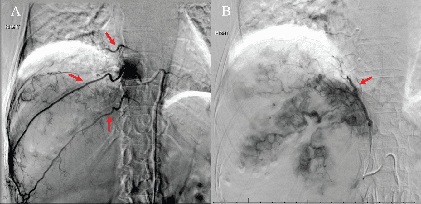
Figure 1. Digital subtraction angiography showing extrahepatic collateral feeding arteries to the hepatic tumor. (A) Selective angiography demonstrating the right tenth and eleventh intercostal arteries (red arrows) supplying the tumor. (B) Selective angiography of the right inferior phrenic artery (red arrow) showing prominent tumor vascularity and staining.
Immediately following the procedure, the patient developed acute right lower-extremity weakness and numbness without bowel or bladder dysfunction. Upon admission, vital signs were notable for a blood pressure of 175/75 mmHg, a heart rate of 54 beats/min, a respiratory rate of 20 breaths/min, a temperature of 36.3°C, and oxygen saturation of 97% on room air. General physical examination revealed mild right upper quadrant abdominal tenderness and intact bilateral pedal pulses. Mental status, speech, and cranial nerve examinations were normal. Neurological examination demonstrated a right T10 sensory level to light touch and temperature, alongside impaired vibratory sensation in the right lower extremity. Left lower-extremity strength was fully preserved (5/5), whereas the right lower extremity exhibited severe paresis: 1/5 in hip flexion, knee flexion, knee extension, and ankle dorsiflexion; 2/5 in hip abduction, hip adduction, foot inversion, and plantar flexion; and 4/5 in hip extension. Deep tendon reflexes were asymmetrically brisk (right patellar 3+, right Achilles 4+ with sustained clonus, and two beats of left ankle clonus), accompanied by bilateral extensor plantar responses.
Postprocedural laboratory evaluation showed elevated transaminases and alkaline phosphatase (alanine aminotransferase 181 U/L, aspartate aminotransferase 178 U/L, and alkaline phosphatase 128 U/L), with unremarkable complete blood counts and serum electrolytes. Non-contrast head CT and brain magnetic resonance imaging (MRI) excluded acute intracranial ischemia or hemorrhage, and CT angiography of the abdomen, pelvis, and bilateral lower extremities ruled out retroperitoneal hematoma, femoral access-site injury, or peripheral arterial occlusion.
Although the initial thoracic spine MRI interpretation was unremarkable, a subsequent neuroradiology addendum identified faint longitudinal hyperintensity on T2-weighted and short tau inversion recovery (STIR) sequences at the T9–T10 and T11–T12 cord levels, without canal or foraminal stenosis (Fig. 2A). Axial T2-weighted images confirmed an intramedullary signal abnormality (Fig. 2B), though fine cross-sectional delineation was partially limited by motion artifact, and diffusion-weighted imaging (DWI) sequences were not obtained. Given the segmental concordance between the embolized intercostal vessels and the neurological deficit, a clinical diagnosis of procedure-associated acute thoracic spinal cord ischemia was established. The patient was initiated on aspirin (325 mg loading dose, followed by 81 mg daily). Right lower-extremity motor function showed minimal recovery over a three-day hospitalization, and she was discharged to a skilled nursing facility for intensive physical rehabilitation.
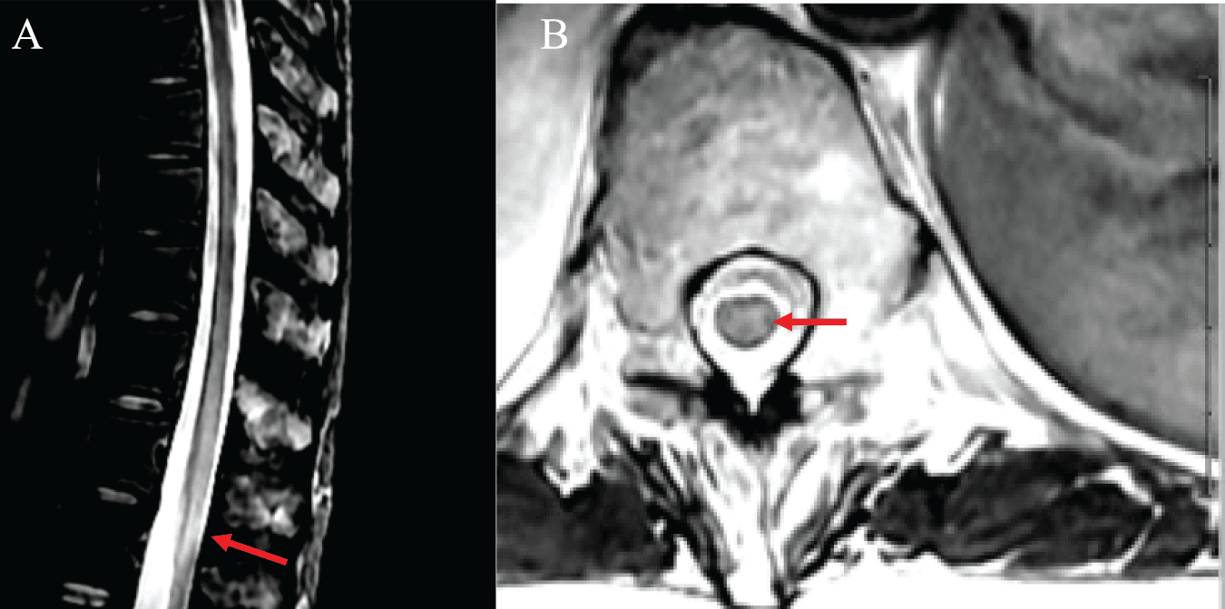
Figure 2. Thoracic spine magnetic resonance imaging suggestive of acute thoracic spinal cord ischemia. (A) Sagittal short tau inversion recovery image showing faint longitudinal hyperintensity in the lower thoracic spinal cord at T9–T10 and T11–T12 (red arrow). (B) Axial T2-weighted image demonstrating intramedullary high signal intensity within the spinal cord (red arrow), with finer evaluation partially limited by motion artifact.
Discussion
The abrupt onset of unilateral upper motor neuron signs, a right T10 spinothalamic sensory level, and ipsilateral vibratory loss immediately after the intervention localizes the lesion to an asymmetric lower thoracic myelopathy, rather than a femoral access-site or peripheral nerve injury. Notably, the initial thoracic spine MRI was interpreted as normal, and only a subsequent specialist addendum identified the faint T2-weighted and STIR hyperintensities at T9–T10 and T11–T12, corresponding to the embolized ninth through eleventh intercostal levels. This diagnostic trajectory highlights an important clinical lesson: because conventional T2 and STIR sequences can appear subtly abnormal or falsely reassuring in hyperacute spinal cord ischemia, especially when axial images are degraded by motion artifact and DWI/apparent diffusion coefficient (ADC) sequences are unavailable, rigorous bedside neurological localization is essential to prompt targeted neuroradiological re-evaluation.6 Even so, the preserved contralateral motor strength, concurrent posterior column involvement, and imaging limitations preclude definitive assignment to a single classic spinal arterial territory, rendering procedure-associated spinal cord ischemia a strongly suspected rather than angiographically proven diagnosis.
Thoracolumbar vascular anatomy explains the potential hazard of embolizing lower intercostal and diaphragmatic tumor feeders. The artery of Adamkiewicz, the dominant anterior radiculomedullary feeder that reinforces the anterior spinal artery, arises between T8 and L1 in approximately 89% of individuals; although 76.6% originate on the left, right-sided origins occur in nearly one-quarter of cases.7 Because the treated right ninth through eleventh intercostal arteries lie directly within this anatomical window, a connection to the spinal circulation is anatomically plausible, and fine radiculomedullary branches can remain unopacified on routine angiography or C-arm CT unless specifically sought.8 Additionally, the RIPA frequently supplies posterior or diaphragmatic HCCs and shares established anastomoses with lower posterior intercostal branches.9 Embolization across these territories can therefore threaten spinal cord perfusion either directly via an unrecognized intercostal radiculomedullary feeder or indirectly across phrenic–intercostal collaterals, even when neither the AKA nor a patient-specific spinal pathway is visualized intraprocedurally.
Two nonexclusive mechanisms may account for spinal cord ischemia in this setting: proximal interruption of segmental arterial inflow to a radiculomedullary branch, and inadvertent distal penetration or reflux of embolic material into spinal feeders.5 The relative likelihood of these mechanisms depends on the specific embolic agent, particle size, liquid concentration, catheter-tip position, and injection dynamics. In our patient, embolization of the right ninth through eleventh intercostal arteries and the right inferior phrenic artery preceded the acute onset of neurological deficits. Both interruption of radiculomedullary arterial inflow and inadvertent migration of embolic material into spinal arterial branches represent plausible mechanisms. Although the temporal relationship and anatomical distribution support procedure-associated spinal cord ischemia, the precise vascular pathway remains uncertain without direct angiographic confirmation.
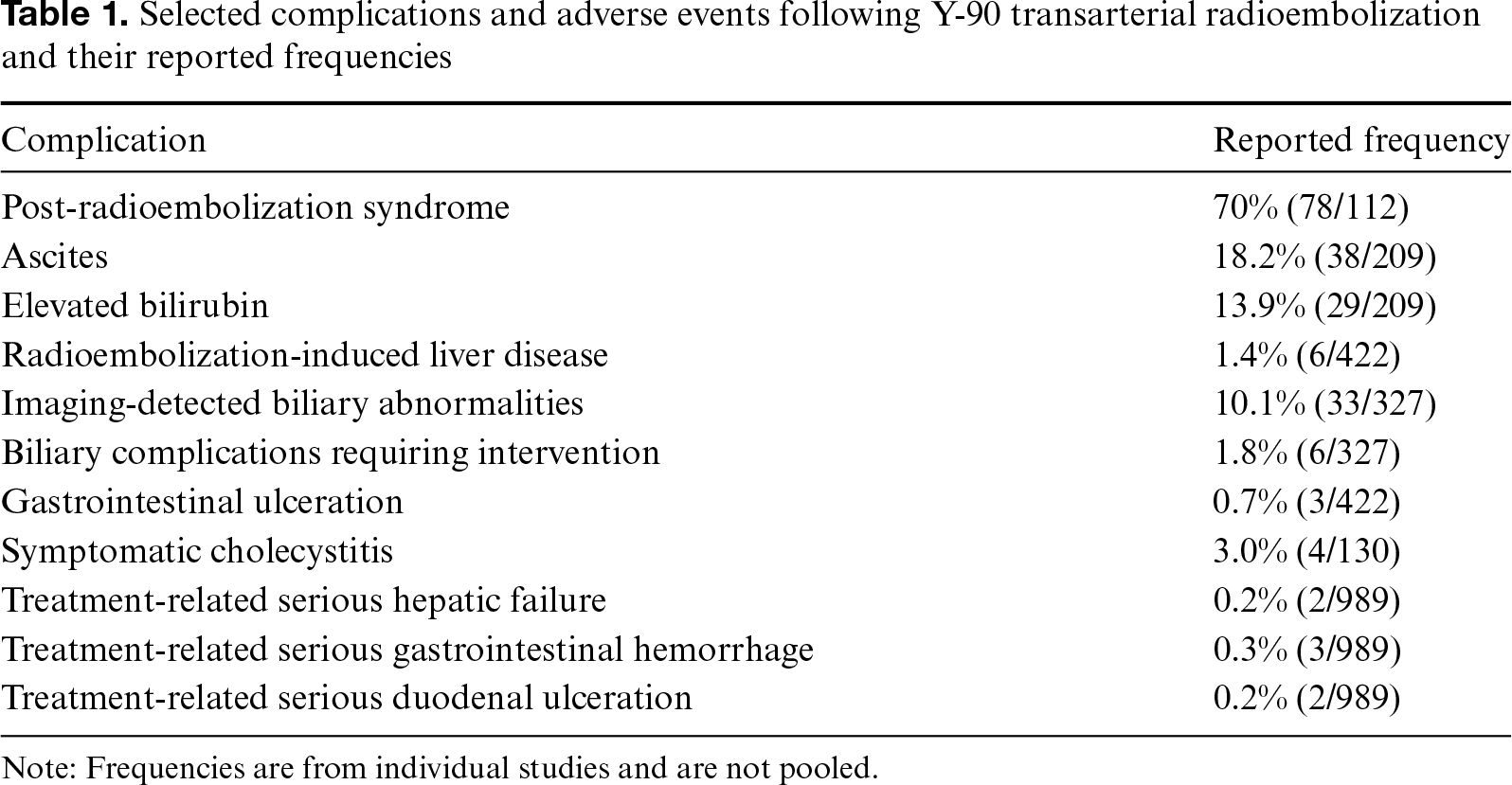
It is clinically important to distinguish adjunctive extrahepatic bland or liquid embolization from intrahepatic Y-90 microsphere delivery. Although glass and resin Y-90 microspheres differ in specific activity and embolic particle burden,10 well-characterized radioembolization complications primarily involve hepatobiliary, gastrointestinal, or systemic toxicities (Table 1).11–15 In this case, particulate and liquid embolic agents were used to embolize the right lower intercostal arteries and the right inferior phrenic artery, followed by administration of glass Y-90 microspheres through right hepatic arterial branches. Given the potential connections between these extrahepatic arteries and the spinal circulation, the neurological complication was more plausibly associated with adjunctive arterial embolization than with intrahepatic Y-90 administration, although the precise vascular mechanism remains uncertain. This case therefore highlights the importance of carefully evaluating selective angiography and C-arm CT for potential spinal arterial branches and tailoring embolization techniques to minimize reflux and nontarget ischemia.
Conclusion
Acute focal neurological deficits developed following adjunctive particle and liquid embolization of the right lower intercostal arteries and the right inferior phrenic artery during a Y-90 TARE treatment course. Prompt bedside neurological localization is vital when early conventional spine MRI appears normal. A careful evaluation for potential spinal arterial connections and a clear distinction between extrahepatic embolization risks and intrahepatic Y-90 toxicities remain essential.
Acknowledgment
The authors sincerely thank the patient for providing informed consent for the publication of this case report.
Funding
This study received no external funding.
Author Contributions
K.L.U., A.H., A.L., and G.C. contributed to the preparation and critical revision of the manuscript. All authors reviewed and approved the final version of the manuscript and agreed to be accountable for its content.
Data Availability Statement
The data supporting the findings of this case report are available from the corresponding author upon reasonable request, subject to patient confidentiality and institutional policies.
Ethics Statement
Written informed consent for publication of this case report was obtained from the patient.
Conflicts of Interest
The authors declare no conflicts of interest.
Generative AI Declaration
Generative AI was not used in the preparation of the manuscript.
Supplemental Information
Supplemental information for this article can be found online at https://sup.jclinque.com/api/articles/130/download-suppl.
References
[1] Sung H, Filho AM, Laversanne M, et al. Global cancer statistics 2024: GLOBOCAN estimates of incidence and mortality worldwide for 34 cancers in 186 countries. CA Cancer J Clin. July–August 2026;76(4):e70090. doi:10.3322/caac.70090.
[2] Sangro B, Argemi J, Ronot M, et al. EASL Clinical Practice Guidelines on the management of hepatocellular carcinoma. J Hepatol. February 2025;82(2):315–374. doi:10.1016/j.jhep.2024.08.028.
[3] Fite EL, Makary MS. Advances and emerging techniques in Y-90 radioembolization for hepatocellular carcinoma. Cancers (Basel). April 2025;29 17(9):1494. doi:10.3390/cancers17091494.
[4] Abdelmaksoud MH, Louie JD, Kothary N, et al. Embolization of parasitized extrahepatic arteries to reestablish intrahepatic arterial supply to tumors before yttrium-90 radioembolization. J Vasc Interv Radiol. October 2011;22(10):1355–1362. doi:10.1016/j.jvir.2011.06.007.
[5] Semba R, Katsumori T, Miura H. Intercostal artery embolization and the risk of spinal cord ischemia: importance of identifying the Adamkiewicz artery-A report of two cases. Radiol Case Rep. September 2026;21(9):3731–3734. doi:10.1016/j.radcr.2026.05.040.
[6] Zedde M, De Falco A, Zanferrari C, et al. Spinal cord infarction: clinical and neuroradiological clues of a rare stroke subtype. J Clin Med. February 2025;15 14(4):1293. doi:10.3390/jcm14041293.
[7] Taterra D, Skinningsrud B, Pękala PA, et al. Artery of Adamkiewicz: a meta-analysis of anatomical characteristics. Neuroradiology. August 2019;61(8):869–880. doi:10.1007/s00234-019-02207-y.
[8] Hieu LT, Van Thanh L, Van Quang V, Khue DK, Anh NHN, Tien DD. Spinal cord ischemia after transcatheter artery chemoembolization for hepatocellular carcinoma: A case-report. Int J Surg Case Rep. May 2023;106(5):108258. doi:10.1016/j.ijscr.2023.108258.
[9] Miyayama S, Matsui O, Taki K, et al. Extrahepatic blood supply to hepatocellular carcinoma: angiographic demonstration and transcatheter arterial chemoembolization. Cardiovasc Intervent Radiol. January-February 2006;29(1):39–48. doi:10.1007/s00270-004-0287-y.
[10] Busse NC, Al-Ghazi M, Abi-Jaoudeh N, et al. AAPM medical physics practice guideline 14.a: Yttrium-90 microsphere radioembolization. J Appl Clin Med Phys. February 2024;25(2):e14157. doi:10.1002/acm2.14157.
[11] Peterson JL, Vallow LA, Johnson DW, et al. Complications after 90Y microsphere radioembolization for unresectable hepatic tumors: An evaluation of 112 patients. Brachytherapy. November–December 2013;12(6):573–579. doi:10.1016/j.brachy.2013.05.008.
[12] Lam M, Garin E, Maccauro M, et al. A global evaluation of advanced dosimetry in transarterial radioembolization of hepatocellular carcinoma with Yttrium-90: the TARGET study. Eur J Nucl Med Mol Imaging. August 2022;49(10):3340–3352. doi:10.1007/s00259-022-05774-0.
[13] Kolligs F, Arnold D, Golfieri R, et al. Factors impacting survival after transarterial radioembolization in patients with hepatocellular carcinoma: Results from the prospective CIRT study. JHEP Rep. February 2023;5(2):100633. doi:10.1016/j.jhepr.2022.100633.
[14] Atassi B, Bangash AK, Lewandowski RJ, et al. Biliary sequelae following radioembolization with Yttrium-90 microspheres. J Vasc Interv Radiol. May 2008;19(5):691–697. doi:10.1016/j.jvir.2008.01.003.
[15] Damm R, Bregenzer C, Steffen I, et al. Cholecystitis induced by Yttrium-90 radioembolization of advanced liver tumors: prospective evaluation of a management algorithm in 197 consecutive patients. Acta Radiol. October 2023;64(10):2783–2790. doi:10.1177/02841851231189490.
 |
Copyright: © 2026 Uy et al. This work is licensed under a Creative Commons Attribution 4.0 International License, which permits unrestricted use, distribution, and reproduction in any medium, provided the original work is properly cited. |